Electrical Signals In Neurons
All living cells have a separation of charges across the cell membrane. This separation of charges gives rise to the resting membrane potential .
Myelin, a fatty insulating material derived from the cell membranes of glial cells, covers the axons of many vertebrate neurons and speeds the conduction of action potentials. The importance of this myelin covering to normal nervous system function is made painfully obvious in individuals with demyelinating diseases in which the myelin covering of the axons is destroyed. Among these diseases is multiple sclerosis, a demyelinating disease of the central nervous system that can have devastating consequences, including visual, sensory, and motor disturbances.
Although neurons share many of the features found in other cell types, they have some special characteristics. For example, neurons have a very high metabolic rate and must have a constant supply of oxygen and glucose to survive. Also, mature neurons lose the ability to divide by . Until the late twentieth century it was thought that no new neurons were produced in the adult human brain. However, there is evidence that, at least in some brain areas, new neurons are produced in adulthood. This finding suggests an exciting avenue for possible approaches to treating such common neurological diseases as Parkinson’s disease and Alzheimer Disease, which are characterized by the loss of neurons in certain brain areas.
SEE ALSO
How Motor Neurons Work In Practice
Think of the process of standing up from a chair. Your brain tells the motor neurons in your legs to activate. Next your motor neurons send instructions to the muscles controlling your legs to rise up. Finally, you might press your arms against the arms of the chair to provide an additional lift.
This series of movements is entirely controlled by the activity of motor neurons. Impressively, it can all happen without much thought at all. The motor neurons work in concert with your muscles to move the body seamlessly through space.
Conduction Of Nerve Impulse
A nerve impulse is the electric signals that pass along the dendrites to generate a nerve impulse or an action potential. An action potential is due to the movement of ions in and out of the cell. It specifically involves sodium and potassium ions. They are moved in and out of the cell through sodium and potassium channels and sodium-potassium pump.
Conduction of nerve impulse occurs due to the presence of active and electronic potentials along the conductors. Transmission of signals internally between the cells is achieved through a synapse. Nerve conductors comprise relatively higher membrane resistance and low axial resistance. The electrical synapse has its application in escape reflexes, heart and in the retina of vertebrates. They are mainly used whenever there is a requirement of fast response and timing being crucial. The ionic currents pass through the two cell membrane when the action potential reaches the stage of such synapse.
Don’t Miss: Get The Message Algebra With Pizzazz
Difference Between Neurons And Neuroglia
The nervous system of higher vertebrates is made of two types of cells namely neuroglia and neurons .
Neurons have two types of processes, viz, dendrons and axons, and forms synapse. On the other hand, neuroglia possesses only one type of process and do not from synapse. Majority of neurons are not capable of differentiating and multiplying in the mature nervous system whereas neuroglia can multiply themselves.
Read on to explore the important difference between neurons and neuroglia in detail.
Following are the important difference between neurons and neuroglia:
| Neurons | |
| They receive and transmit nerve impulses | They provide mechanical as well as structural support to the neurons |
| Granules | |
| Around 5-10 times the neurons in higher vertebrates | |
| Age & number | |
| The number does not change with age | The number decreases with age |
| Size | |
| 4 micrometer to 1 millimeter | Smaller than neurons |
| It involves itself in signal transduction | Supplies nutrients to neurons |
| The functional unit of the nervous system | They are supporting cells of neurons |
What Is Neuronal Cell Biology

Neuronal cell biology falls under the larger umbrella of cellular biology, the realm of science that pokes, prods, and illuminates the most basic unit of life. Compared to other cell types, however, the neurons inner workings are not as well understood. The mystery stems in part from the very nature of nerve cells: theyre large and highly polarized, meaning one end the tree-like dendrites looks and functions very differently than the other end a long, cord-like projection called an axon. In addition, the electrical environment inside the cell is vastly different from the outside, a contrast that facilitates the propagation of electrical signals along the axon. And while the cells unique morphology can prove challenging for experimentalists in the lab, the same qualities facilitate graceful cell-to-cell communication in the brain.
Read the News Release:
Also Check: How Did The Geography Of Greece Affect Its Development
Function Of Sensory Neurons
Sensory neurons make up all the senses in the body, even those of which you are not consciously aware! The function of sensory neurons is to detect and transmit signals from a peripheral region to a more central location in the central nervous system, i.e., the spinal cord or the brain.
The transduction of the signal takes place in the sensory receptor at the dendritic end of the neuron. This is where the new signal is generated in response to a stimulus, such as a smell, touch, or taste.
The stimulus triggers the sensory neuron to send a signal then carries information towards the central nervous system. Specifically, depolarization is initiated at the sensory receptors and transmitted along the dendrites to the cell body and then to the axon. At the axon terminal, the signal initiates the release of chemicals into the synapse. These chemicals are what trigger the response in the spinal cord.
Neurons And Glial Cells
The information below was adapted from OpenStax Biology 35.1 and Khan Academy AP Biology The neuron and nervous system. All Khan Academy content is available for free at www.khanacademy.org
The nervous system is made up of neurons, the specialized cells that can receive and transmit chemical or electrical signals, and glia, the cells that provide support functions for the neurons. A neuron can be compared to an electrical wire: it transmits a signal from one place to another. Glia can be compared to the workers at the electric company who make sure wires go to the right places, maintain the wires, and take down wires that are broken. Recent evidence suggests that glia may also assist in some of the signaling functions of neurons.
Neurons communicate via both electrical signals and chemical signals. The electrical signals are action potentials, which transmit the information from one of a neuron to the other the chemical signals are neurotransmitters, which transmit the information from one neuron to the next. An action potential is a rapid, temporary change in membrane potential , and it is caused by sodium rushing to a neuron and potassium rushing out. Neurotransmitters are chemical messengers which are released from one neuron as a result of an action potential they cause a rapid, temporary change in the membrane potential of the adjacent neuron to initiate an action potential in that neuron.
Also Check: How To Calculate A Half Life
No Service: What Is Neuronal Signalling
When you think of signal, you think of that ancient technology you used to use to text people and have to hang out of your window to stop your call breaking up. Now, the word signal is normally replaced with data or Wi-Fi, but both of the latter are still types of signals. They allow you to receive information and send out responses in the blink of an eye. So how do neurons communicate?
Well, the technology a neuron uses to signal may not appear to be quite as fancy as the new IoS or windows update, but the principals are similar to the very basic code these softwares are built on. Neurons communicate using an all or none signal called an action potential. At the synapse, action potentials are passed from one neuron to another. These signals are sometimes described as electric signals as they use ions inside and outside the neuron for their conduction, similar to electrical conduction.
The all-or-none nature of the action potential means the neuron either sends a full action potential or nothing at all. There is no half or small action potential each one is the exact same size. This is similar to binary code . Everything on your screen right now is coded by a series of 1s and 0s and everything in your head is coded by action potentials or not . When an action potential is triggered, there is a short period when no other signal can be transmitted. This is known as the refractory period and prevents constant action potential signalling.
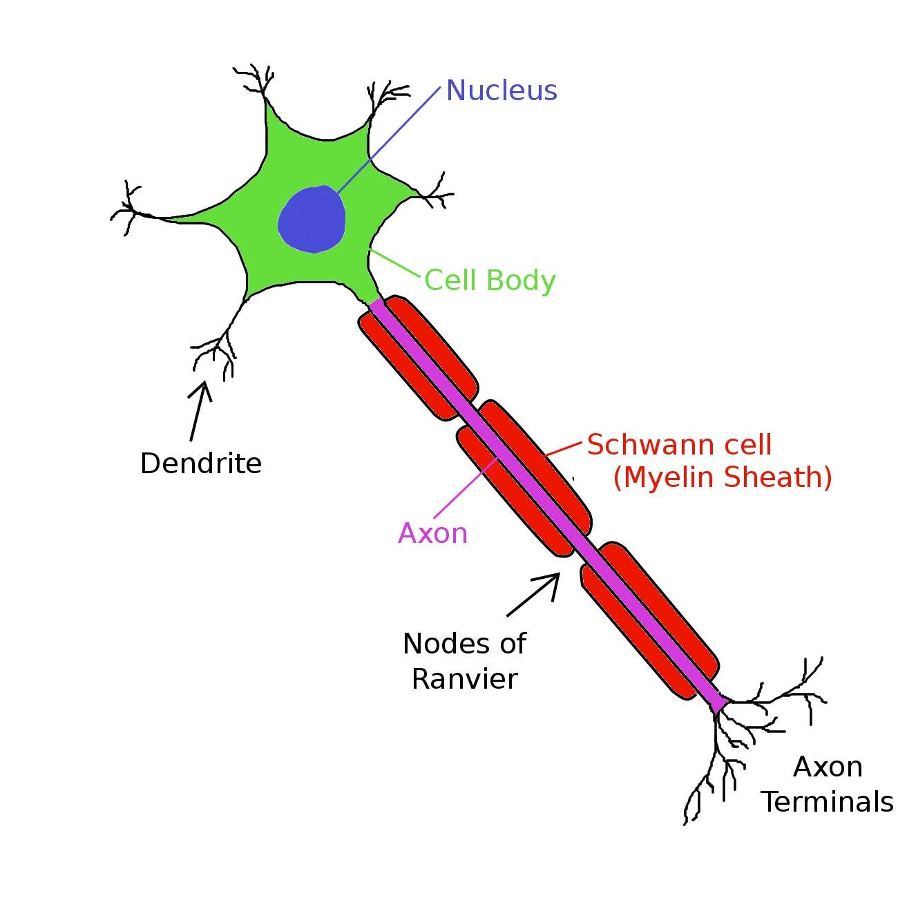
The Parts Of A Neuron:
- Dendrites: these are the points of connections between neurons . When a connection is made with the soma of another cell , this is a stronger connection.
- Soma: The cell body of a neuron. This is the part of the neuron that is most like other cells. It has the nucleus, genetic machinery and is where many of the metabolic processes happen.
- Axon: This is where the magic happens. The Axon has a base where it is continuous with the soma. This swollen section is called the axon hillock, or the spike initiating zone . The nerve impulse then travels down the axon to the Axon Terminals.
- Axon Terminals: These are the ends of axons. They contain neurotransmitters. When the nerve impulse reaches the axon terminals, the axon terminals release the neurotransmitter, which may then result in an action potential in the next cell.
There are three basic types of neurons: Sensory neurons, motor neurons and interneurons. Sensory neurons bring signals to the central nervous system and Motor neurons send signals from the CNS to the rest of the body. Interneurons are in the CNS and allow the sensory neurons to communicate with the motor neurons.
The take home message is that neurons are what its all about in the nervous system.
You May Like: Rationalizing Imaginary Denominators Worksheet Answers
Histology And Internal Structure
Numerous microscopic clumps called Nissl bodies are seen when nerve cell bodies are stained with a basophilic dye. These structures consist of rough endoplasmic reticulum and associated ribosomal RNA. Named after German psychiatrist and neuropathologist Franz Nissl , they are involved in protein synthesis and their prominence can be explained by the fact that nerve cells are very metabolically active. Basophilic dyes such as aniline or haematoxylin highlight negatively charged components, and so bind to the phosphate backbone of the ribosomal RNA.
The cell body of a neuron is supported by a complex mesh of structural proteins called neurofilaments, which together with neurotubules are assembled into larger neurofibrils. Some neurons also contain pigment granules, such as neuromelanin , and lipofuscin , both of which accumulate with age. Other structural proteins that are important for neuronal function are actin and the tubulin of microtubules. Class III β-tubulin is found almost exclusively in neurons. Actin is predominately found at the tips of axons and dendrites during neuronal development. There the actin dynamics can be modulated via an interplay with microtubule.
There are different internal structural characteristics between axons and dendrites. Typical axons almost never contain ribosomes, except some in the initial segment. Dendrites contain granular endoplasmic reticulum or ribosomes, in diminishing amounts as the distance from the cell body increases.
Direction
Structure Of Sensory Neurons
A typical neuron is comprised of dendrites, an axon, and a cell body, and the sensory neurons are no exception. Most sensory neurons are pseudounipolar, which means they have a single axon extending from the cell body that forms two extensions: the dendrites and the axon.
The sensory neuron begins with the dendrites, as this is where the signal is received from the external environment. The dendrites branch-like projections that receive the signal. In sensory neurons, the dendrites are generally very long.
The signal from the receptors at the end of the dendrites travels along the nerve fiber until it reaches the cell body. The cell body is the location of the nucleus, cytoplasm, and other organelles. In pseudounipolar sensory neurons, the cell body is located in a side branch of the nerve fiber.
The fiber that carries the signal away from the cell body is called the axon. In sensory neurons, the axon is short. The axon communicates with the spinal cord, and the end of the axon is where the neuron terminates.
The nerve fibers are protected, insulated, and nourished by a fatty cushion, called a myelin sheath. The myelin sheath is made of a layer of Schwann cells.
Don’t Miss: 4.5 Practice B Geometry Answers
Show/hide Words To Know
Axon: a long thick projection in nerve cells that sends electrical signals out away from the cell body……more
Dendrites: long thin projections in nerve cells which receive electrical signals… more
Myelin sheath: covers the axon and works like insulation to help keep electrical signals inside the cell, making them move more quickly.
Nerve: many neurons bundled together.
Neuron: a special cell which is part of the nervous system. Neurons work together with other cells to pass chemical and electrical signals throughout the body… more
Nucleus: where DNA stays in the cell, plural is nuclei.
Soma: the main cell body.
Synapse: a gap between two cells that lets chemical or electrical signals be passed between them… more
Unique: one of a kind.
Sensory Motor And Interneurons

Neurons are also categorized according to the target tissues they communicate with. Sensory neurons are primarily pseudo-unipolar and found in both peripheral and central nervous tissue. They pick up information according to the five senses smell, taste, hearing, sight, and touch, as well as data that helps us to know how our body is positioned and balanced .
Spinal motor neurons are also part of the central nervous system . Upper motor neurons travel between the brain and spinal cord, and lower motor neurons between the spinal cord and muscle. A lower motor neuron always ends at a muscle fiber and is predominantly multipolar.
Interneurons or relay neurons connect a sensory nerve cell to a motor nerve cell as shown in the neuron diagram below. There can be more than one interneuron in a circuit.
Interneurons are found in the spinal cord where they play a significant role in reflex arcs. In the cortex of the brain, interneurons seem to connect multiple regions to provide a complete set of patterns or behaviors it has been noted that many psychiatric disorders show abnormal interneuron function. Current research has only managed to scrape the surface of the different cortical interneurons and we still have much to learn.
In the cerebellum, interneurons such as granule cells and unipolar brush cells regulate other functional neurons such as Purkinje cells that inhibit the firing rate of other neurons.
Also Check: Mcdougal Geometry Workbook Answers
Educators And Parents Sign Up For The Cheat Sheet
Weekly updates to help you use Science News for Students in the learning environment
Thank you for signing up!
There was a problem signing you up.
What makes these cells special
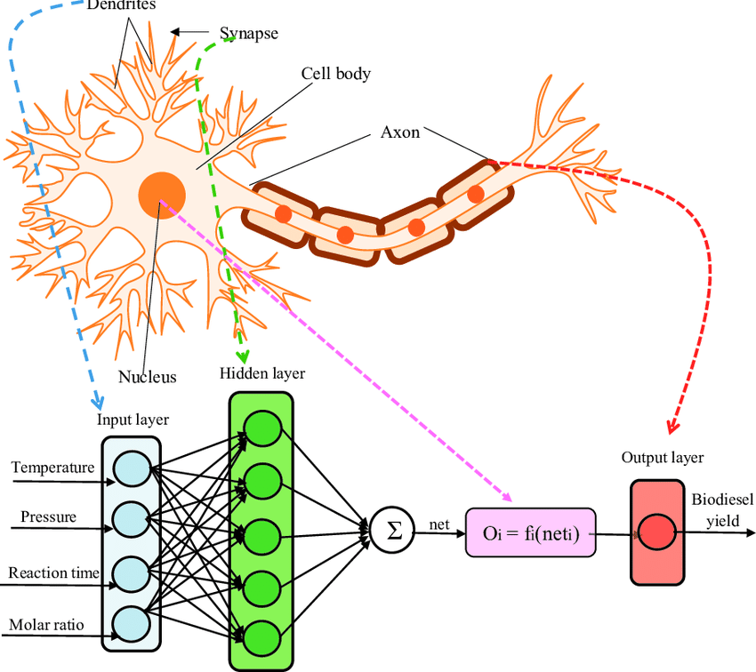
Neurons have all of the basic features of animal cells. For instance, they have a nucleus and an outer membrane. But unlike other cells, they also have branching hair-like structures called dendrites. These catch chemical messages from other cells. The dendrites send each impulse to the main part of the cell. Its known as the cell body. From there, the signal moves along a long thin section of the cell called the axon. This electrical impulse is made by waves of charged particles weaving in and out of the cell membrane, rippling the signal along. Some axons have fatty rings of myelin on them, lined up like beads on a string. When the neurons are myelinated, the message will bounce along much faster.
The message leaves an axon through finger-like terminals at the end. Chemicals released out of the cell here will then be picked up by the dendrites on a neighboring cell. The area from one cells terminals, across the gap between cells and on to the next cells dendrites is known as a synapse . Messages pass between one cell and onto the next by floating across the space between a gap called the synaptic cleft. This tiny space between the two cells is filled with fluid. In the next neuron, the chemical signals enter molecules called receptors like a key into a lock.
Anatomy of a Neuron
Neurons Vs Other Cells
-
Neurons and other body cells both contain a nucleus that holds genetic information.
-
Neurons and other body cells are surrounded by a membrane that protects the cell.
-
The cell bodies of both cell types contain organelles that support the life of the cell, including mitochondria, Golgi bodies, and cytoplasm.
-
Neurons stop reproducing shortly after birth. Generally, when neurons die they are not replaced, although neurogenesis, or the formation of new nerve cells, does occur in some parts of the brain.
-
Research has shown that new connections between neurons form throughout life.
-
Neurons have a membrane featuring an axon and dendrites, specialized structures designed to transmit and receive information.
-
Neurons release chemicals known as neurotransmitters into synapses, or the connections between cells, to communicate with other neurons.
Don’t Miss: What Is Elastic Force
Diffuse Patterns Of Synaptic Connections Are Sharpened By Activity
In a normal animal the retinotectal map is initially fuzzy and imprecise: the system of matching markers we have just described is enough to define the broad layout of the map, but not sufficient to specify its fine details. Studies in frogs and fish show that each retinal at first branches widely in the tectum and makes a profusion of synapses, distributed over a large area of tectum that overlaps with the territories innervated by other axons. These territories are subsequently trimmed back by selective elimination of synapses and retraction of axon branches. This is accompanied by the formation of new sprouts, through which each axon develops a denser distribution of synapses in the territory that it retains.
A central part in this remodeling and refinement of the map is played by two competition rules that jointly help to create spatial order: axons from separate regions of retina, which tend to be excited at different times, compete to dominate the available tectal territory, but axons from neighboring sites in the retina, which tend to be excited at the same time, innervate neighboring territories in the tectum because they collaborate to retain and strengthen their synapses on shared tectal cells . The mechanism underlying both these rules depends on electrical activity and signaling at the synapses that are formed. If all action potentials are blocked by a toxin that binds to voltage-gated Na+ channels, remodeling is inhibited and the map remains fuzzy.