What Is Cytosis Medical
4.6/5cytosis
Similarly, you may ask, what is the definition of Cytosis in biology?
Cytosis is a transport mechanism for the movement of large quantities of molecules into and out of cells. There are three main types of cytosis: endocytosis , exocytosis , and transcytosis .
Subsequently, question is, what suffix means blood? emia: Suffix meaning blood or referring to the presence of a substance in the blood. As for example, anemia and hypervolemia .
Simply so, what does Cyto mean in medical terms?
Cyto-: Prefix denoting a cell. “Cyto-” is derived from the Greek “kytos” meaning “hollow, as a cell or container.” From the same root come the combining form “-cyto-” and the suffix “-cyte” which similarly denote a cell.
What does philia mean in medical terms?
–philia. , -phily , -philous. Combining forms meaning that which has an attraction to or is stained by.
In Summary: Endocytosis And Exocytosis
Cells perform three main types of endocytosis. Phagocytosis is the process by which cells ingest large particles, including other cells, by enclosing the particles in an extension of the cell membrane and budding off a new vesicle. During pinocytosis, cells take in molecules such as water from the extracellular fluid. Finally, receptor-mediated endocytosis is a targeted version of endocytosis where receptor proteins in the plasma membrane ensure only specific, targeted substances are brought into the cell.
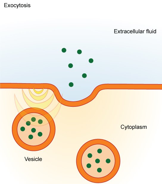
Exocytosis in many ways is the reverse process from endocytosis. Here cells expel material through the fusion of vesicles with the plasma membrane and subsequent dumping of their content into the extracellular fluid.
What Is Endocytosis Endocytosis Definition And Purposes
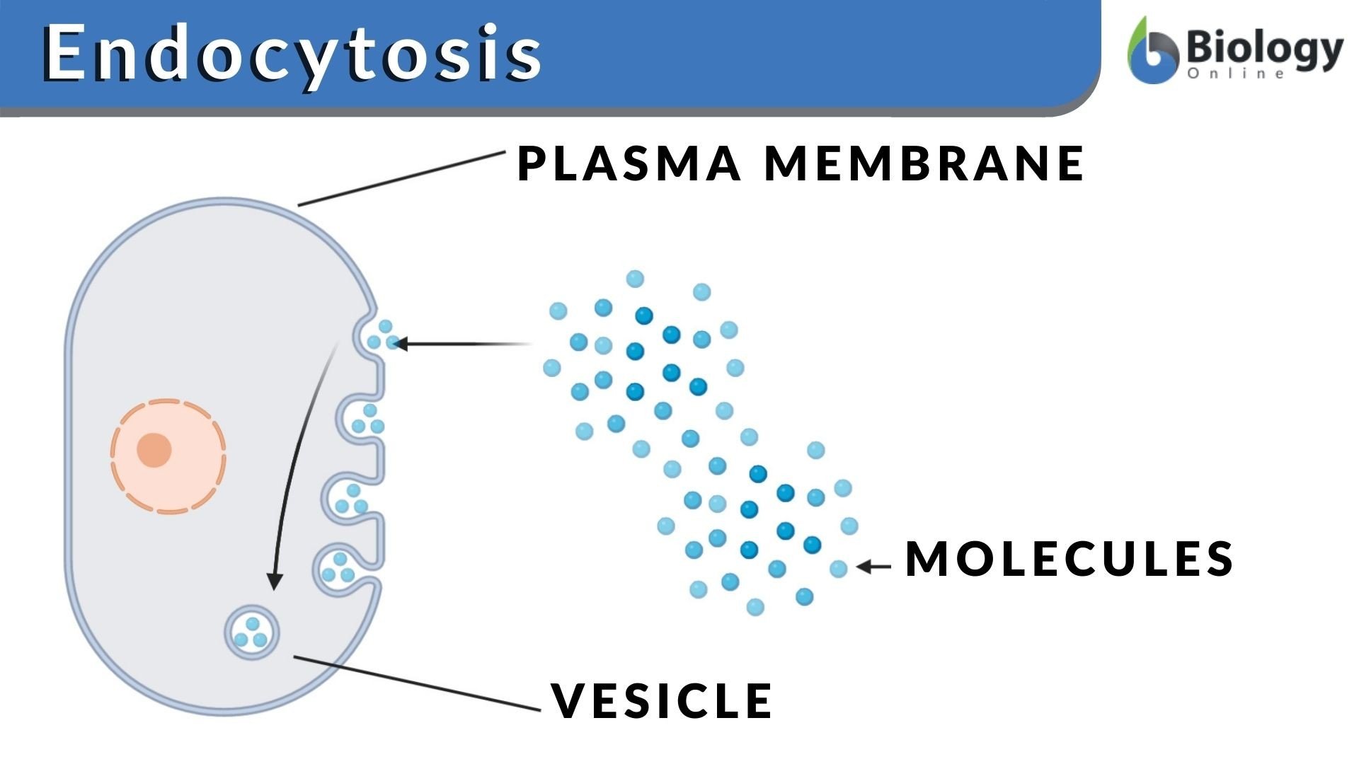
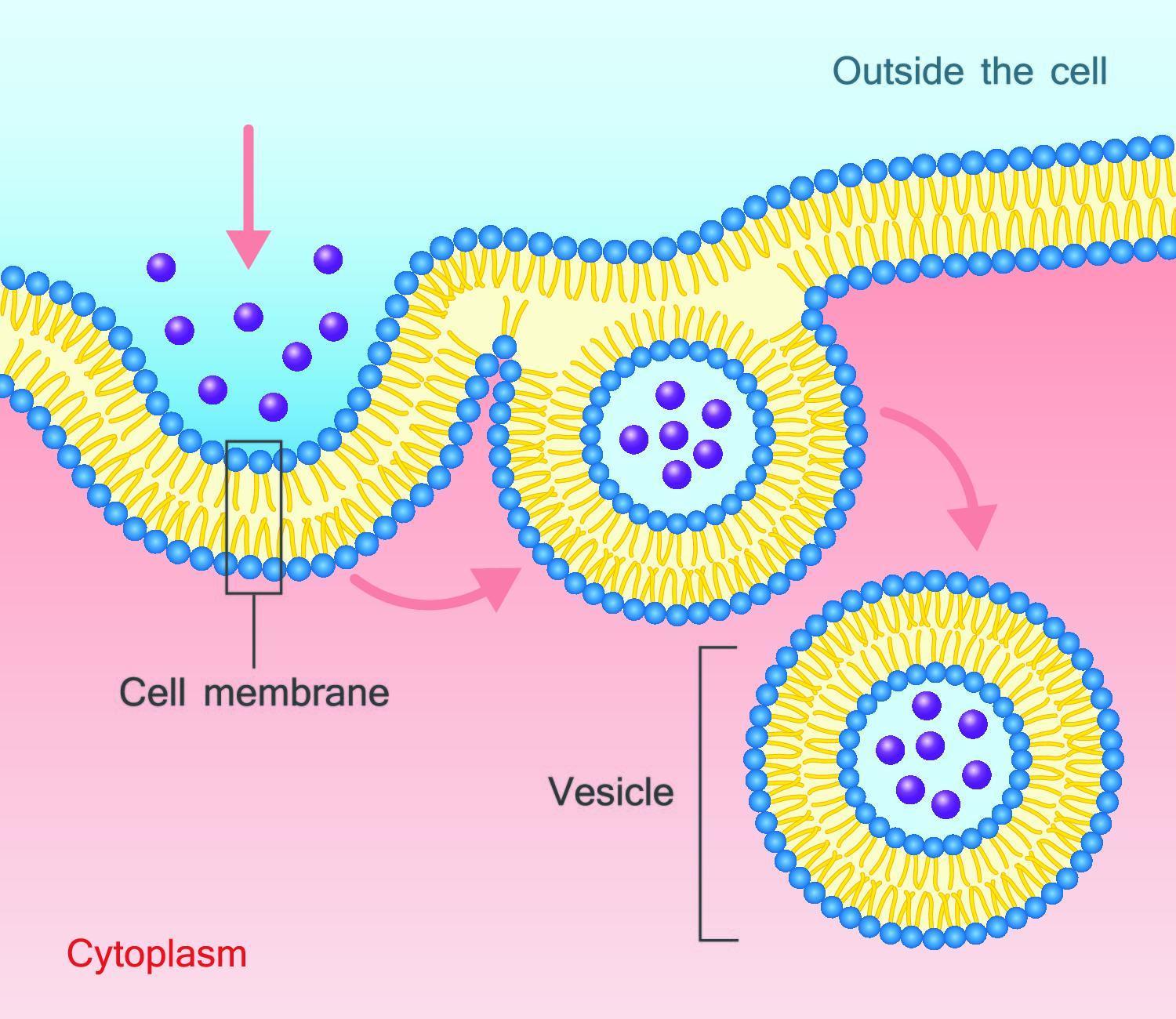
Endocytosis is the process by which cells take in substances from outside of the cell by engulfing them in a vesicle. These can include things like nutrients to support the cell or pathogens that immune cells engulf and destroy.Endocytosis occurs when a portion of the cell membrane folds in on itself, encircling extracellular fluid and various molecules or microorganisms. The resulting vesicle breaks off and is transported within the cell.Endocytosis serves many purposes, including:
- Taking in nutrients for cellular growth, function and repair: Cells need materials like proteins and lipids to function.
- Capturing pathogens or other unknown substances that may endanger the organism: When pathogens like bacteria are identified by the immune system, they are engulfed by immune cells to be destroyed.
- Disposing of old or damaged cells: Cells must be safely disposed of when they stop functioning properly to prevent damage to other cells. These cells are eliminated through endocytosis.
Don’t Miss: Prentice Hall Gold Algebra 1 Teaching Resources Answers Chapter 9
Describe The Primary Mechanisms By Which Cells Import And Export Macromolecules
In addition to moving small ions and molecules through the membrane, cells also need to remove and take in larger molecules and particles. Some cells are even capable of engulfing entire unicellular microorganisms. You might have correctly hypothesized that the uptake and release of large particles by the cell requires energy. A large particle, however, cannot pass through the membrane, even with energy supplied by the cell.
There are two primary mechanisms that transport these large particles: endocytosis and exocytosis.
Vesicle Function In Endocytosis And Exocytosis

During bulk transport, larger substances or large packages of small molecules are transported through the cell membrane, also known as the plasma membrane, by way of vesicles think of vesicles as little membrane sacs that can fuse with the cell membrane.Cell membranes are comprised of a lipid bilayer. The walls of vesicles are also made up of a lipid bilayer, which is why they are capable of fusing with the cell membrane. This fusion between vesicles and the plasma membrane facilitates bulk transport both into and out of the cell.
Recommended Reading: Definition Of Span Linear Algebra
Main Difference Endocytosis Vs Phagocytosis
Endocytosis and phagocytosis are two mechanisms involved in taking in material into the cell. Endocytosis contains two categories: phagocytosis and pinocytosis. This matter that is taken into the cell could be enzymes, hormones, nutrients, ions, cell debris, dead cells or even bacteria like pathogens inside the body of a multicellular organism. Both endocytosis and phagocytosis form vesicles, surrounding the uptaken matter. Exocytosis is involved in the elimination of waste substances, which are produced by the digestion inside these vesicles. The main difference between endocytosis and phagocytosis is that endocytosis is taking in of matter into a living cell by forming vesicle by the cell membrane whereas phagocytosis is taking in of large solid matter into the cell by forming phagosomes.
This article looks at,
3. What is the difference between Endocytosis and Phagocytosis
Endocytic Pathways: Different Entry Portals And Sorting Routes
Endocytosis occurs by distinct mechanisms in mammalian cells . A traditional way to differentiate between the different endocytic pathways is on the basis of cargo size. Particles larger than 500 nm are taken up by phagocytosis, a specific form of endocytosis that is dependent on the receptorligand interaction and occurs in specialized cells, such as immune system cells, which can actively remove solid particles, for example, pathogens and cell debris .110 Another endocytic pathway relies on macropinocytosis, which is responsible for the uptake of smaller particles as well as of fluids and solutes.111 Macropinocytosis is able to internalize cargoes that are up to 500 nm in diameter, which subsequently fuse with lysosomes to hydrolyze the cargo particles. Both phagocytosis and macropinocytosis involve large rearrangements of the PM, guided by extensive actin cytoskeleton remodeling and coordinated by the stepwise involvement of Rho-GTPases.112,113
CME and NCE will be discussed in detail in the following sections.
L. Aurelian, in, 2014
You May Like: Tenmarks Answer Hack
What Is Pinocytosis In Biology
biologypinocytosispinocytosis
What is the function of pinocytosis?
Pinocytosis, a process by which liquid droplets are ingested by living cells. Pinocytosisis one type of endocytosis, the general process by which cells engulf external substances, gathering them into special membrane-bound vesicles contained within the cell.
what is phagocytosis in biology?
Contents
What Is Pinocytosis Example
Examples of Pinocytosis Cells in the kidney can use pinocytosis to separate nutrients and fluids from the urine that will be expelled from the body. In addition, human egg cells also use it to absorb nutrients prior to being fertilized.
What is Pinocytosis and phagocytosis?
Pinocytosis and Phagocytosis are the two categories of Endocytosis. Phagocytosis is an intake of solid particles with the formation of vesicles called phagosomes, while pinocytosis is the intake of liquid particles with the formation of vesicles called pinosomes.
You May Like: Paris Jackson Biological Parents
Exocytosis In The Pancreas
Exocytosis is used by a number of cells in the body as a means of transporting proteins and for cell to cell communication. In the pancreas, small clusters of cells called islets of Langerhans produce the hormones insulin and glucagon. These hormones are stored in secretory granules and released by exocytosis when signals are received.
When glucose concentration in the blood is too high, insulin is released from islet beta cells causing cells and tissues to take up glucose from the blood. When glucose concentrations are low, glucagon is secreted from islet alpha cells. This causes the liver to convert stored glycogen to glucose. Glucose is then released into the blood causing blood-glucose levels to rise. In addition to hormones, the pancreas also secretes digestive enzymes by exocytosis.
Endocytosis And Exocytosis: Differences And Similarities
Complete the form below and we will email you a PDF version of“Endocytosis and Exocytosis: Differences and Similarities”
First Name*Would you like to receive further email communication from Technology Networks?Technology Networks Ltd. needs the contact information you provide to us to contact you about our products and services. You may unsubscribe from these communications at any time. For information on how to unsubscribe, as well as our privacy practices and commitment to protecting your privacy, check out our Privacy Policy
Endocytosis and exocytosis are the processes by which cells move materials into or out of the cell that are too large to directly pass through the lipid bilayer of the cell membrane. Large molecules, microorganisms and waste products are some of the substances moved through the cell membrane via exocytosis and endocytosis.
Also Check: Segment Addition Postulate Kuta
The Steps Of Endocytosis
The following is an outline of the basic steps of the two types of endocytosis.
Two types of endocytosis: phagocytosis and pinocytosis.
Phagocytosis:
Pinocytosis:
Molecular Mechanisms Of Endocytosis

- Philip G WoodmanAffiliationsDivision of Biochemistry, School of Biological Sciences, University of Manchester, Oxford Road, Manchester, M13 9PT, United Kingdom
- Gerrit van MeerAffiliationsAcademic Medical Center, University of Amsterdam, Department of Cell Biology and Histology, 1105 AZ, Amsterdam, The Netherlands
You May Like: Eoc Fsa Practice Test Algebra 2 Calculator Portion
Endocytosis In Signaling And Development
In addition to its well-described roles in nutrient uptake and receptor down-regulation, endocytosis plays a direct role in the modulation of cell signaling responses . Marcos Gonzalez-Gaitán presented studies on Smad anchor for receptor activation, SARA, which links Dpp receptors with Smad signaling adaptor proteins and is a central component of the Dpp signaling pathway in Drosophila. SARA is located to a subset of endosomes that mediate cellular memory of Dpp signaling in wing imaginal discs by distributing evenly between the two daughter cells during cell division . Using development of the sensory organ precursor as a model for asymmetrical cell division, Gonzalez-Gaitán showed that SARA-containing endosomes accumulate at the central spindle during cell division and then partition asymmetrically into the signal-receiving daughter cell. Even though SARA is a mediator of Dpp signaling, the SARA-containing endosomes could well function as vehicles for other signaling pathways, and Gonzalez-Gaitán is currently investigating the possibility that asymmetrical Notch-Delta signaling, strongly implicated in binary fate decisions, could be mediated via SARA-containing endosomes. Another evidence that endocytosis regulates SOP development was provided by Roland Le Borgne , who proposed that transcytosis of Delta may be required for Notch-Delta signaling in the SOP.
Endocytic Routes Specify Signaling Outcome
Figure 2. Different endocytosis routes lead to different biological outputs. EGFR and TGFR can both be endocytosed via CME or NCE routes. Receptors endocytosed via CME are recycled back to the plasma membrane thus sustaining the signal, while receptors trafficked via the NME route are directed to lysosomes for degradation. In the case of TGFR, CME to EEA1 positive endosomes leads to receptor coupling with a SMAD anchor protein, SARA, which promotes TGFR signaling from endosomes, whereas the caveolin-dependent internalization pathway leads to rapid receptor turnover.
It is important to note that NCE does not necessarily lead to receptor degradation and CME to endosomal signaling. Wnts constitute a large family of secreted ligands implicated in a wide array of developmental and physiological processes . Wnt-induced low-density receptor-related protein 6 signaling results in NCE and relocalization of LRP6 to endosomes, where the Wnt-generated signal functions to stabilize -catenin . In contrast, in response to Dickkopf family member 1 , a soluble ligand and a negative regulator of Wnt signaling, LRP6 undergoes CME and receptor degradation. In conclusion, the internalization routes of receptors can play a defining role on the signaling outcome upon ligand stimulation and interplay between different trafficking pathways facilitates fine-tuning of cellular signals.
Recommended Reading: What Does Similar Figures Mean In Math
Exocytosis Definition: What Is Exocytosis
Exocytosis is a form of active transport that allows cells to move large molecules into the extracellular membrane, where they can be utilized in various ways. This is a critical process for all living things, from plants and invertebrates to protozoa and human beings. As mentioned, this is a form of active transport, rather than passive, because the majority of the large molecules being made by cells are polar in nature, meaning that they cannot pass through the cell membrane without assistance, due to the hydrophobic aspect of the membrane.
When a cell is ready to move a molecule into the outer environment, the molecule is bound in a secretory vesicle, which can be thought of as a suitcase for complex molecules. This vesicle will then move to the outer edge of the cell and temporarily fuse with the membrane. At that point, the water-soluble molecule inside the vesicle can be dumped out into the extracellular environment.
There are two main types of exocytosis: Ca2+ triggered non-constitutive exocytosis and non-Ca2+ triggered constitutive exocytosis. The difference is that in the former, the process is more regulated, requiring direct external signals to occur, an increase in calcium within the cell, and a clathrin coat on the vesicle. This is the type of exocytosis that occurs between synapses and neurons. All other forms of exocytosis are simpler and less regulated, despite still being considered a form of active transport and requiring the expenditure of ATP.
The Basic Steps Of Endocytosis
There are three primary types of endocytosis: phagocytosis, pinocytosis, and receptor-mediated endocytosis. Phagocytosis is also called “cell eating” and involves the intake of solid material or food particles. Pinocytosis, also called “cell drinking”, involves the intake of molecules dissolved in fluid. Receptor-mediated endocytosis involves the intake of molecules based upon their interaction with receptors on a cell’s surface.
Also Check: Operations With Complex Numbers Kuta Software
Roles In Virus Biology
Glycoproteins, Gn and Gc, play a significant role in the biology of hantaviruses, including virus entry to host cells, virulence, and assembly and packaging of new virions in infected cells.
aAttachment and entry
Hantaviruses enter the cells via receptor-mediated endocytosis. The acidic environment of the endosome facilitates fusion between the viral and cellular membranes, with a consequent release of the viral nucleocapsids into the cytoplasm . Viral glycoproteins mediate attachment of virus to endothelial cells via interaction with surface 3 integrin receptors .
bAssembly and packaging
cVirulence
Martina Rieder, Karl-Klaus Conzelmann, in, 2011
What Happens After Exocytosis
In most cases, the secretory vesicle cannot be recovered in its full form to be reused, as it has bonded with the cell membrane. The creation of vesicles is an energy-intensive process, and requires significant effort by the mitochondria, the energy factories in cells, to carry out this process. In non-constitutive exocytosis, such as neuronal exocytosis, where neurotransmitters are transported, the vesicles are not reused.
That being said, there are some forms of vesicle fusion that are not permanent, such as kiss and run fusion, which allows a partial deposit of vesicle contents, and then a reformation of the vesicle membrane. Recent research has also identified partial emptying of vesicles in some cases, where a secretory vesicle will release some of its contents, detach and reseal, and float back into the intracellular space. It can then return to the membrane and release the rest of its contents, or be re-filled with a new molecule at a receptor site for another organelle.
Don’t Miss: Segment Addition Postulate Unit 1 Geometry Basics
Systems Biology Approaches To Study Endocytosis
Current technologies enable global analyses of physiological pathways, and a few systems biology approaches to study endocytosis in various organisms were presented. Genome-wide analysis of endocytic recycling in S. cerevisiae was reported by Liz Conibear . The screen involved measuring an increased or decreased presence of the v-SNARE Snc1p on the plasma membrane, thus identifying both exocytic and endocytic defects among the mutant collection encompassing deletions of all nonessential genes. Genetic interaction analysis of the top hits established a network of gene clusters involved in various intracellular processes contributing to Snc1p trafficking, containing both known and novel components.
Why Is Bulk Transport Important For Cells

Cell membranes are semi-permeable, meaning they allow certain small molecules and ions to passively diffuse through them. Other small molecules are able to make their way into or out of the cell through carrier proteins or channels.But there are materials that are too large to pass through the cell membrane using these methods. There are times when a cell will need to engulf a bacterium or release a hormone. It is during these instances that bulk transport mechanisms are needed.Endocytosis and exocytosis are the bulk transport mechanisms used in eukaryotes. As these transport processes require energy, they are known as active transport processes.
Recommended Reading: Holt Geometry Chapter 7 Test Form B Answer Key
Role Of Phosphorylation In Exocytosis And Endocytosis Of Aquaporin
Although S256 phosphorylation is necessary for vasopressin-induced cell-surface accumulation of AQP2, the role that phosphorylation plays in AQP2 exocytosis is complex. An AQP2 S256A mutant accumulates on the plasma membrane upon inhibition of endocytosis ,253 suggesting that the exocytotic pathway is intact under these conditions. Vasopressin also increases exocytosis of vesicles in AQP2-expressing cells whether or not AQP2 is phosphorylated at S256.344 Thus, although vasopressin-induced accumulation of AQP2 at the cell surface requires S256 phosphorylation, and AQP2 is present in endocytosis-resistant membrane domains after vasopressin treatment,253,345,346 exocytotic insertion of AQP2 into the plasma membrane is probably independent of this phosphorylation event. Furthermore, the regulated endocytosis of AQP2 may not be dependent on its phosphorylation state.347 For example, prostaglandin E2 348 can induce AQP2 internalization independent of S256 phosphorylation, but other studies indicate that the effects of PGE2 on AQP2 and urine concentration depend on which PGE2 receptor it acts upon.109,250,344,349351
J. Alanko, … J. Ivaska, in, 2016