What Is An Eeg Test
What to expect when undergoing this test
By Olivia Guy-Evans, published June 16, 2021
The electroencephalogram is a neuroimaging testwhich can detect and record minute changes in electrical activity within the brain. This is recordedusingmacroelectrodes.
It produces a chart which shows howâbrain wavesâ vary byfrequency andamplitude of electrical output from thebrain changes over time.
German physiologist Hans Berger was known as the inventor of the EEG back in 1924 where he conducted the first EEG on a human.
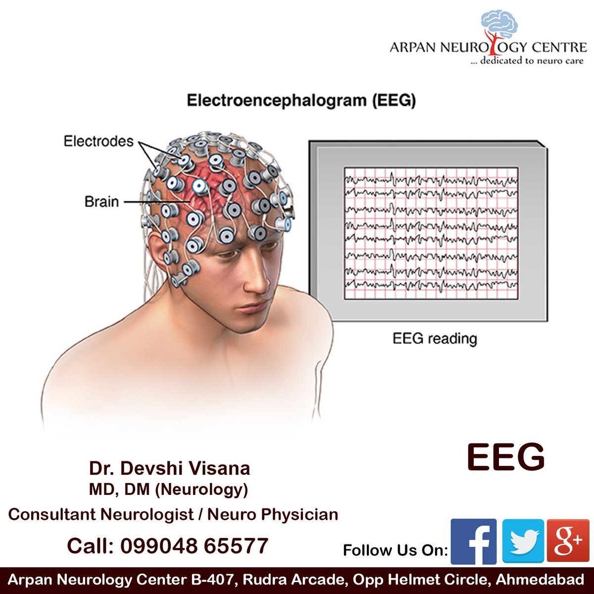
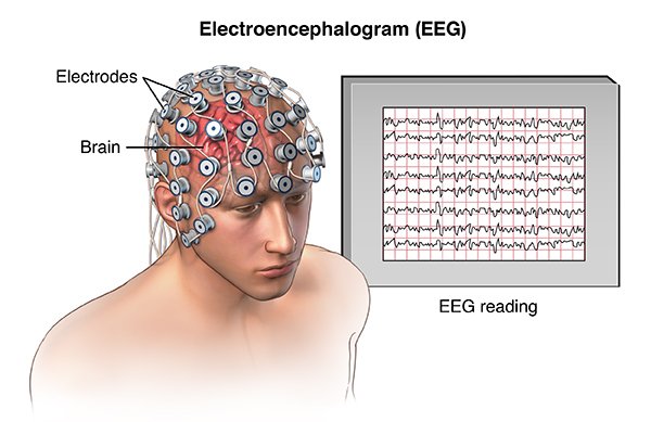
The cells of the brain communicate to each other via electrical impulses, which are picked up by the EEG through small metal discs, called electrodes, which get attached to the scalp.
The electrodes cannot pick up signals for individual neurons. The recording shows the electrical activity from small areas of the brain. EEG is used to show the presence or absence of specific brain activity in specific areas of the brain, with an accuracy within milliseconds.
The electrodes of the EEG analyze the electrical impulses that are being communicated in the brain, which then gets transmitted to a computer which monitors the results.
The electrical activity in the brain is known as action potential, or nerve impulses. This is how neurons pass messages to each other in order for behaviors, thought, and other conscious or unconscious processes to occur.
Eeg Vs Fmri Fnirs Fus And Pet
EEG has several strong points as a tool for exploring brain activity. EEGs can detect changes over milliseconds, which is excellent considering an action potential takes approximately 0.5â130 milliseconds to propagate across a single neuron, depending on the type of neuron. Other methods of looking at brain activity, such as PET, fMRI or fUS have time resolution between seconds and minutes. EEG measures the brain’s electrical activity directly, while other methods record changes in blood flow or metabolic activity , which are indirect markers of brain electrical activity.
EEG can be used simultaneously with fMRI or fUS so that high-temporal-resolution data can be recorded at the same time as high-spatial-resolution data, however, since the data derived from each occurs over a different time course, the data sets do not necessarily represent exactly the same brain activity. There are technical difficulties associated with combining EEG and fMRI including the need to remove the MRI gradient artifact present during MRI acquisition. Furthermore, currents can be induced in moving EEG electrode wires due to the magnetic field of the MRI.
EEG can be used simultaneously with NIRS or fUS without major technical difficulties. There is no influence of these modalities on each other and a combined measurement can give useful information about electrical activity as well as hemodynamics at medium spatial resolution.
Does Emotiv Offer Eeg Monitoring Solutions
EMOTIVs solutions have been proven in studies and clinical literature for neuroscience, workplace wellness and safety, cognitive performance, neuromarketing, and brain-controlled technology applications.
EMOTIV offers three different headsets which can be used for EEG monitoring:
The award-winning EMOTIV EPOC+ EEG headset provides professional-grade brain data for academic research within biometrics research and commercial use. The EMOTIV Insight headset boasts minimal set-up time and electronics optimized to produce clean signals from anywhere, making it ideal for performance and wellness tracking. The EMOTIV EPOC FLEX cap offers high-density coverage and movable electroencephalogram sensors optimal for research professionals. EmotivPRO is an integrated brain research software solution for neuroscience research and education, built for EPOC+, EPOC Flex, and Insight headsets.
You May Like: Who Are Paris Jackson’s Biological Parents
Medical Issues To Consider
An abnormal EEG doesn’t automatically mean that you, for example, have epilepsy. The EEGs of babies and young children can often record irregular patterns that don’t mean anything, or the irregularities may flag previously diagnosed neurological conditions such as cerebral palsy. On the other hand, a normal EEG doesn’t rule out epilepsy either. Sometimes, a person with epilepsy will only display abnormal brain waves during a seizure.
How Do I Get Ready For An Eeg

When preparing to have an EEG, it is usually advisable to wash your hair the night before or on the day of the test, and to not use conditioners. This is because hair products can make it more difficult for the electrodes to adhere to the scalp.
It is also advisable to not consume anything with caffeine or alcohol as this may affect the results.
Typically, you should be allowed to take your usual medications unless otherwise instructed by the doctor.
If you are supposed to be sleeping during your EEG test, your doctor may advise you to not sleep the night before, or to sleep less, so that you are able to take part in the test.
Read Also: What Is The Molecular Geometry Of Ccl4
How Does An Eeg Machine Work
The EEG electrodes pick up on electrical activity produced by neurons. EEG machines use an array of electrodes because the brain produces different signals from different brain regions. The number of electrodes corresponds with the number of channels an EEG machine has. The more channels, the higher the resolution of EEG data captured. A 32 channel EEG machine captures a more detailed picture of brainwave activity than an 8 channel EEG machine, in other words.
EEG signals are typically very small around 10 microVolts or less. To make accurate measurements the signals from the electrodes are passed to an amplifier system that stabilizes the signals and magnifies them to a level that can be measured accurately using common electronic components that convert them to digital signals. The amplified signals can be recorded via computer, mobile device or cloud database.
Are There Any Risks Or Side Effects
The EEG procedure is painless, comfortable and generally very safe. No electricity is put into your body while it’s carried out. Apart from having messy hair and possibly feeling a bit tired, you normally will not experience any side effects.
However, you may feel lightheaded and notice a tingling in your lips and fingers for a few minutes during the hyperventilation part of the test. Some people develop a mild rash where the electrodes were attached.
If you have epilepsy, there’s a very small risk you could have a seizure while the test is carried out, but you’ll be closely monitored and help will be on hand in case this happens.
Read Also: Eoc Algebra 1 Practice Test 2015
Eeg In Research Applications
In addition to its diagnostic potential, EEG has tremendous research value. Indeed, the technology has been used to explore brain function for nearly a century, and has been applied across diverse corners of psychology and neuroscience. Cognitive psychologists, for instance, frequently use EEG to investigate neural correlates of basic cognitive functions, such as emotion, language, attention, and learning. Likewise, some social psychologists use EEG results to augment analysis of group behavior and social cognition.
Increasingly, researchers are looking to EEG not just to diagnose disorders, but to restore function in individuals suffering from paralysis or neurodegenerative disease, or to enhance existing human capabilities. This can be achieved via what is known as a brain-computer interface, or BCI, which translates the brains electrical signals into action. EEG BCIs create a direct connection between the brain and some sort of external device, such as a computer or a robotic arm, granting new levels of control to paralyzed users. Further, there exists substantial momentum in the field of recreational BCIs, which would allow healthy users to control a computer screen using thought alone.
Are There Risks Associated With An Eeg
There are no risks associated with an EEG. The test is painless and safe.
Some EEGs do not include lights or other stimuli. If an EEG does not produce any abnormalities, stimuli such as strobe lights, or rapid breathing may be added to help induce any abnormalities.
When someone has epilepsy or another seizure disorder, the stimuli presented during the test may cause a seizure. The technician performing the EEG is trained to safely manage any situation that might occur.
Recommended Reading: What Is The Molecular Geometry Of Ccl4
How Does Eeg Work
In EEG, an array of flat metal electrodes are placed on a persons scalp. These electrodes measure electrical signals that result from the activity of neurons in the cerebral cortex. The readings can provide a picture of voltage change on the scalp over time, including at intervals shorter than one second. EEG measures can be used as indicators of mental processes or states and can help diagnose conditions such as epilepsy or a sleep disorder. The record of brain activity produced by EEG is called the electroencephalogram.
Immediately After The Eeg
Once the test is complete, the electrodes are removed and you are allowed to get up. The results need to be analysed at a later stage by a neurologist .Generally, if there is no abnormality to the brain’s electrical activity, the pattern of ‘peaks and valleys’ charted by the EEG should be fairly regular. If excited, the pattern will show considerable variation, and any departure from the regular pattern can indicate abnormalities.
Read Also: How Did Geography Discourage Greek Unity
What Is An Eeg Machine
EEG machines which include portable EEG machines, ambulatory EEG machines and EEG neurofeedback machines or EEG biofeedback machines are monitoring devices used for EEG recordings. EEG stands for electroencephalography, the non-invasive method of monitoring the brains electrical signals.
Most of these devices contain electrodes, amplifiers, filters and an analog to digital converter. Wireless or portable EEG machines contain a battery, while wired EEG machines will be hooked up directly to a computer.
An ambulatory EEG machine is used during an extended EEG reading. Often used to diagnose sleep disorders or seizure disorders, ambulatory EEGs record for up to 72 hours while traditional EEG tests record for 1-2 hours.
EEG neurofeedback machines, also called EEG biofeedback machines, are EEG systems that allow a subject to see their brain activity in real-time. During this type of EEG monitoring, the EEG machine is synced to a computer or a cloud device, and the subjects brain waves are displayed on a computer screen.
Eeg Data And Analysis

EEG data analysis can admittedly be a complex process, which is why iMotions has several features designed to reduce the burden of this step.
Frontal alpha asymmetry, a measure used as a proxy for feelings of approach or avoidance, is typically used to provide an assessment of how appealing or repellent a stimulus is. This and power spectral density can be automatically calculated in iMotions, and the R code used to build the analysis is fully available and transparent.
Other manufacturers, such as ABM and Emotiv may also provide the capability to calculate proprietary metrics such as levels of drowsiness or engagement. These metrics are also provided within the iMotions software, giving you easy access to detailed insights.
There may also be parts of the analysis which you want to exclude, or look at in more detail. iMotions provides an annotation tool that can be used either live as data-collection occurs, or following data collection. Its straightforward to mark up the data and select specific segments for processing or export.
The data, whether raw, processed, or segmented can of course also be exported in easily transferable formats, allowing you to take your analysis to whichever platform you prefer. There is also information about computer use, such as mouse clicks and keystrokes, particularly useful when relating stimulus interaction to biosensor data.
Don’t Miss: Which Founding Contributors To Psychology Helped Develop Behaviorism
How Does An Eeg Work
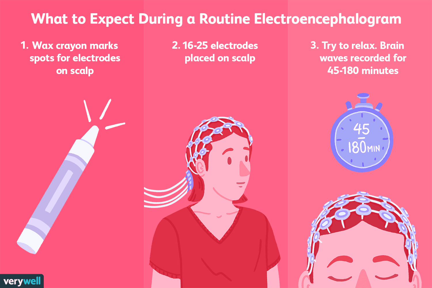
The billions of nerve cells in your brain produce very small electrical signals that form patterns called brain waves. During an EEG, small electrodes and wires are attached to your head. The electrodes detect your brain waves and the EEG machine amplifies the signals and records them in a wave pattern on graph paper or a computer screen .
There are several different ways to conduct an EEG:
- Standard EEG recording is done in the office and usually lasts an hour. You may be asked to do a sleep-deprived EEG, which requires you to have only 4 hours of sleep. Abnormal brain waves may appear when the body is stressed or fatigued. This exam usually takes 2 to 3 hours. You will be given specific instructions regarding food, drink, and medications that may need to be avoided.
- Ambulatory EEG involves wearing a portable EEG recorder on a belt around your waist for several days or weeks. The EEG recorder along with a diary you keep of daily activities and drug dosages helps the doctor relate your activity to specific EEG recordings.
- Video EEG monitoring is available in specialized centers for patients with frequent seizures or sleep disorders. You stay in the hospital and are monitored both by EEG and a video camera. This allows you to be observed during a seizure so that your physical behavior can be monitored at the same time as your EEG.
Which Should You Use
As always, this depends on your research question. If you are more concerned with structural and functional detail, then MRI or fMRI could well be your choice if you are able to make the considerable investment required.
For quicker, affordable, and accessible insights about brain function, with a tight temporal resolution, EEG is the method of choice.
If youd like more guidance in deciding on the method of choice for your research, then reach out and talk to our team.
I hope youve found this discussion and comparison of MRI, fMRI, and EEG helpful. If youd like to get an even deeper understanding of EEG, then download our free guide below!
Recommended Reading: Exponential Growth And Decay Common Core Algebra 1 Homework Answers
How Should I Prepare For The Test
- Make sure your hair is clean, freshly washed, and free from any styling products. If you have long hair, do not braid, tie, or pin it up.
- You may eat regular meals, but avoid drinks that contain caffeine for at least 4 hours before the test.
- Do not nap before the test.
- Continue taking your medicine unless your doctor tells you to stop.
What Is A Bold Signal
In fMRI studies, BOLD stands for blood-oxygen-level-dependent. A BOLD signal is a brain imaging signal that is increased or decreased by the level of oxygen in the blood within any given part of the brain. This signal change is possible because the magnetic properties of blood hemoglobin that lacks oxygen differ from those of oxygenated hemoglobin. Since blood-oxygen levels and blood flow are related to the nearby activity of neurons, a BOLD signal is a useful way to gauge changes in neuronal activity in specific brain areas.
Also Check: Which Founding Contributors To Psychology Helped Develop Behaviorism
How To Read An Eeg Monitor
Electroencephalography is a sensitive means to capture brain activity. Signals not made by the brain, known as artifacts, can interfere with capturing brain wave data. Artifacts can include electrical interference and electrode displacement, or muscle and eye movements. Any of these non-brain signals can interfere with data capture and quality. Medical researchers have established a normal baseline for healthy brain activity. An EEG time series that shows variations from that normal baseline can indicate problems, such as epilepsy or another disorder.
Sleep Study For Sleep Disorders
An EEG sleep study or polysomnography test measures body activity in addition to performing a brain scan. An EEG technologist monitors heart rate, breathing and oxygen levels in your blood during an overnight procedure. Polysomnography is mostly used in medical research and as a diagnostic test for sleep disorders.
Also Check: Segment Addition Postulate Practice Answer Key
What Is Eeg Monitoring For Seizures
Various types of EEG monitoring are used to diagnose and understand seizures or prescribe antiepileptic drugs or epilepsy surgery. Video EEG monitoring pairs EEG test data with a video recording of the seizures clinical and behavioral manifestations. Long term EEG monitoring expands the limited time sampling associated with a routine EEG recording, which may only provide a 20- to a 40-minute sample of brain activity. Ambulatory EEG monitoring allows patients to conduct long term EEG monitoring without being restricted to a hospital or health care setting. Continuous EEG monitoring is used in intensive care units to monitor brain waves in comatose and critically ill patients.
What Are The Risks Of An Eeg

EEGs have been in use for many years and are considered a safe procedure, causing no discomfort. As the electrodes do not produce any sensations, there is very little risk, they cause no discomfort, and there is no risk of getting an electric shock.
EEGs are a non-invasive technique, meaning it does not involve any equipment going into the body. This makes EEGs a good choice in research, especially with younger participants.
The need for the participants to remain still are not required as much as with other neuroimaging methods such as with magnetic resonance imaging . Therefore, participants can carry out a variety of tasks or normal ongoing behaviors for an experiment without the worry of data being disrupted due to movements.
Another advantage of EEGs is that it has very high temporal resolution, meaning it can pick up frequencies really quickly, often within a single millisecond.
In comparison to other neuroimaging techniques such as MRI or positron emission tomography , EEGs hold a strong advantage over these in terms of temporal resolution. Similarly, many other neuroimaging methods will record changes in blood flow or metabolic activity, which are indirect markers of brain activity.
EEGs however, can measure the brainâs electrical activity directly, making it more efficient in this sense.
This is why neuroscientists would discuss EEG activity generated at specific electrode locations rather than concluding that a certain part of the brain generated the activity.
You May Like: Kendall Hunt Geometry Answers