Activation Of A Growth Factor Receptor
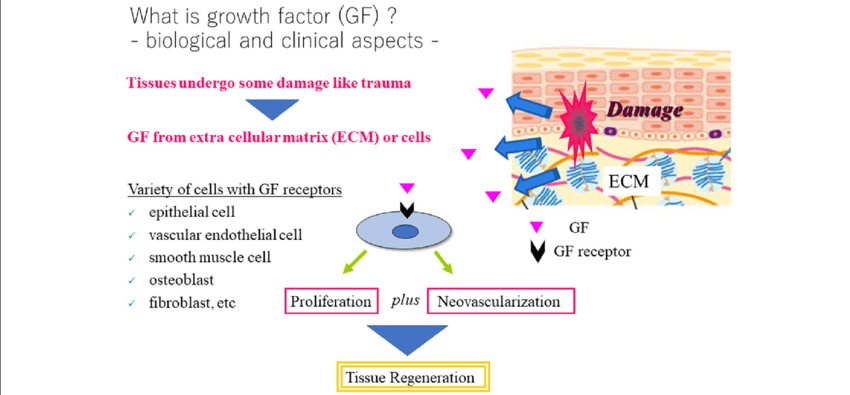
A growth factor receptor is activated by binding to a specific growth at the cell’s surface. The activated receptor, in turn, activates an intracellular protein . A receptor such as platelet-derived growth factor can stimulate a number of substrates including Ras protein, the Src protein , or phospholipase C, a signal transmitter.
AN EXAMPLE… RASPDGF binds to its receptor —-> Ras protein is activated —- > Ras triggers a short, time-limited signal —-> cell division is initiated.
Then, Ras is inactivated by GTPase activation protein .
If a gene mutation is present in either Ras or GAP, the time-limited aspect of the cell-stimulating signals may be lost…resulting in uncontrolled cell division which can lead to tumor formation.
X Therapeutic Applications Of Vegf
Figure 8.Open in new tab
Selective internal iliac angiography of control and VEGF-treated rabbit at day 10 , day 20, and day 40. VEGF was administered as single intraarterial bolus in the iliac artery of rabbits in which the ipsilateral femoral artery had been removed to induce sustained, severe ischemia. Note the modest collateral vessel development in the control, which contrasts with the marked improvement observed after VEGF treatment.
A further potential therapeutic application of VEGF is the prevention of restenosis after percutaneous transluminal angioplasty. Between 15% and 75% of patients undergoing percutaneous transluminal angioplasty for occlusive coronary or peripheral arterial disease develop restenosis within 6 months . It has been proposed that damage to the endothelium is a crucial event triggering fibrocellular intimal proliferation . Therefore, the induction of rapid reendothelialization may be an effective strategy to prevent the cascade of events leading to neointima formation and ultimately to restenosis in patients. Recent evidence shows that VEGF accelerates reendothelialization and also attenuates intimal hyperplasia in balloon-injured rat carotid artery or rabbit aorta .
Current Therapeutic Strategy Targeting Ctgf
Connective tissue growth factor is considered as a therapeutic target to combat cancer, fibrosis and other related disorders in a variety of organs, and tissues. There are many approaches such as antibodies, synthetic peptides, small interfering RNAs , and antisense oligonucleotides , targeting CTGF to exert therapeutic effect .
Table 4. Summary of drugs targeting CTGF.
Don’t Miss: Mcdougal Littell Geometry Practice Workbook Answer Key
New Technology For Drug Discovery Targeting Ctgf
Although antibodies are now established as a key therapeutic modality for a range of diseases, their limited stability, complicated in vivo production, and typically undefined cross-reactivity are challenges to overcome . Aptamers are short single stranded oligonucleotides that bind to their targets through 3D conformational complementarities with high affinity and specificity . The aptamer-based drug Macugen was approved by the Food and Drug Administration in 2004 for the treatment of neovascular age-related macular degeneration and a series of aptamer-based drugs are in clinical pipelines . Aptamers are considered to be strong chemical rivals of antibodies due to their inherent advantages over antibodies. Compared to antibodies: aptamers can be produced using cell-free chemical synthesis and are therefore less expensive to manufacture, aptamers exhibit extremely low variability between batches and have better controlled post-production modification, aptamers are minimally immunogenic, aptamers are more stable at room temperature and longer shelf life, and aptamers are small in size and could bind to regions which are inaccessible to antibodies . Moreover, aptamers could specifically target a specific protein from family proteins with highly similar structures .
Types Of Growth Factors

There are Four Classes of Growth Factors:
- Class I comprises growth factors interacting with specific receptors at the cell surface and includes epidermal growth factor , growth hormone , and platelet- derived growth factor .
- Class II are cell surface hormone receptors, frequently protein tyrosine kinases in the cytoplasmic domains of the receptor, as for EGF and PDGF.
- Class III consists of a large group of intracellular signal transmitters, or transducers, belonging to different families, e.g., Ras proteins and protein kinases such as Src.
- Class IV are nuclear transcription factors . Initiators of DNA transcription include fos, myc, myb, and N-myc. Suppressors of cell division are such proteins as p53 and the retinoblastoma gene product.
Don’t Miss: Half Life Chemistry Equation
Connective Tissue Growth Factor And Ccn Family
CTGF, also known as CCN2, is a 38 kDa, cysteine-rich , extracellular matrix protein that belongs to the CCN family of proteins . The term connective tissue growth factor was introduced to describe a novel polypeptide growth factor that stimulated DNA synthesis and chemotaxis in fibroblast . There are other five CCN gene family genes: CCN1 , CCN3 , CCN4 , CCN5 , and CCN6 . The CCN acronym was introduced from the names of the first three members of the family to be discovered: Cyr61 , CTGF and NOV . Expression of CTGF is crucial to embryonic development in childhood , for example, mice with CTGF knockout have multiple skeletal dysmorphisms and perinatal lethality . Also, abnormal expression of CTGF was detected in several adulthood diseases including fibrosis and malignancy in major organs and tissues .
Modifying Gfs Signaling And Functionality
The signaling properties of GFs can be modified to enhance their regenerative activity. The next section focuses on different approaches that attempt to modify the sequence or the structure of GFs to promote their function , thereby effecting similar or altogether different responses at lower doses. Although those strategies have the potential to produce highly effective modified GFs, they may require longer development as the effects of modified signaling may be less predictable than those of improved delivery or stability.
Don’t Miss: Mega Hack V5 Geometry Dash
Growth Factors Vs Mitogens
According to Campbell Biology,
A growth factor is a protein released by certain cells that stimulates other cells to divide.
and according to Wikipedia,
A mitogen is a chemical substance that encourages a cell to commence cell division, triggering mitosis.
Whats the difference between them? I suppose a mitogen specifically refers to mitosis, but if something is stimulating mitosis, isnt it stimulating growth? Are mitogens all growth factors? Are all growth factors mitogens?
There is a lot of confusion and conflicting / imprecise definitions of these terms. It’s biology after all 🙂
A mitogen is an agent that causes a cell to enter mitosis. This definition is pretty clear, and there is a good consensus about it.
The term growth factor has at least two different definitions: a factor that causes growth of tissues, organs or entire individuals or a factor that causes growth of cells . These two versions are often mixed up, and this causes no end of confusion. Let’s consider them both in turn.
You can find a great article about this here.
basically, these two groups are very similar in their effects, however they work through different pathways.
Growth factors usually stimulates mTOR receptor, which is a “serine/threonine protein kinase that regulates cell growth, cell proliferation, cell motility, cell survival, protein synthesis, autophagy, and transcription.”
I hope this helps.
Engineering Gfs For Non
GFs can be immobilized to the ECM or ECM-derived biomaterials through affinity binding by the introduction of an ECM-binding sequence or domain at either terminus of the GF. The strategy presents the advantage of giving modified GFs the ability to bind the endogenous ECM where the GF is delivered, in some cases allowing to forgo the use of exogenous biomaterials altogether. Such approach allows GFs to be more readily available for resident cells by being immobilized in the local ECM instead of having to be released by biomaterials. In addition, the simplicity of biomaterial-free delivery systems could lead to a higher cost-effectiveness. However, the effectiveness of these strategies may depend on the local ECM composition.
As one of the most abundant ECM proteins, collagens represent good binding targets for engineering GFs for delivery to collagen-rich tissues. For example, a bacterial collagen-binding domain , was fused to fibroblast growth factor-2 , allowing improved bone formation in a spinal fusion model . In another study, CBD-fused FGF-2 showed the ability to induce significantly higher mesenchymal cell proliferation and callus formation in a mice fracture model compared to wild-type FGF-2 . Similarly, a CBD-fused hepatocyte growth factor delivered via hydrogel improved recovery after spinal cord injury in mice compared to wild-type HGF .
Recommended Reading: What Happened To Jonathan Thomas Child Of Rage
Ii Biological Activities Of Vegf
VEGF is a potent mitogen for micro- and macrovascular endothelial cells derived from arteries, veins, and lymphatics, but it is devoid of consistent and appreciable mitogenic activity for other cell types . The denomination of VEGF was proposed to emphasize such narrow target cell specificity . VEGF promotes angiogenesis in tridimensional in vitro models, inducing confluent microvascular endothelial cells to invade collagen gels and form capillary-like structures . These studies provided evidence for a potent synergism between VEGF and bFGF in the induction of this effect . Also, VEGF induced sprouting from rat aortic rings embedded in a collagen gel . This model emphasizes the specificity of VEGF, as the proliferation induced by this growth factor consisted almost exclusively of vascular endothelial cells. In contrast, insulin-like growth factor-I or platelet-derived growth factor induced endothelial cell sprouting accompanied by extensive fibroblastic proliferation . VEGF also elicits a strong angiogenic response in a variety of in vivo models including the chick chorioallantoic membrane , the rabbit cornea , the primate iris , the rabbit bone , etc.
Get The Growth Facts About Growth Factors
To help you get your desired cell culture results every time, weve compiled a list from experienced researchers of important facts about growth factors.
The growth facts
Don’t Miss: Core Connections Algebra Answers
Epidermal Growth Factor Receptor
| EGFR |
|---|
| Ortholog search: PDBeRCSB |
| List of PDB id codes |
| 11 A2|11 9.41 cM | Start |
|---|
| View/Edit Mouse |
The epidermal growth factor receptor is a transmembrane protein that is a receptor for members of the epidermal growth factor family of extracellular protein ligands.
The epidermal growth factor receptor is a member of the ErbB family of receptors, a subfamily of four closely related receptor tyrosine kinases: EGFR , HER2/neu , Her 3 and Her 4 . In many cancer types, mutations affecting EGFR expression or activity could result in cancer.
Epidermal growth factor and its receptor was discovered by Stanley Cohen of Vanderbilt University. Cohen shared the 1986 Nobel Prize in Medicine with Rita Levi-Montalcini for their discovery of growth factors.
Deficient signaling of the EGFR and other receptor tyrosine kinases in humans is associated with diseases such as Alzheimer’s, while over-expression is associated with the development of a wide variety of tumors. Interruption of EGFR signalling, either by blocking EGFR binding sites on the extracellular domain of the receptor or by inhibiting intracellular tyrosine kinase activity, can prevent the growth of EGFR-expressing tumours and improve the patient’s condition.
A Distribution Of Vegf Flk

The proliferation of blood vessels is crucial for a wide variety of physiological processes such as embryonic development, normal growth and differentiation, wound healing, and reproductive functions. Previous studies have indicated that the VEGF mRNA is temporally and spatially related to the proliferation of blood vessels in the rat, mouse, and primate ovary and in the rat uterus, suggesting that VEGF is a mediator of the cyclical growth of blood vessels that occurs in the female reproductive tract . In fact, in situ hybridization studies in the rat ovary provided the first evidence that VEGF may be a regulator of physiological angiogenesis .
It has been suggested that VEGF is also involved in a major pathophysiological process such as wound healing . Keratinocytes in a healing wound express VEGF mRNA. Interestingly, a decreased expression of VEGF mRNA has been observed in the skin of genetically diabetic db/db mice , suggesting that an altered regulation of VEGF gene expression contributes to defective angiogenesis and impaired wound healing characteristic of this disorder.
Recommended Reading: Colorful Overnight Geometry Dash
Growth Factor And Control Of Cell Growth
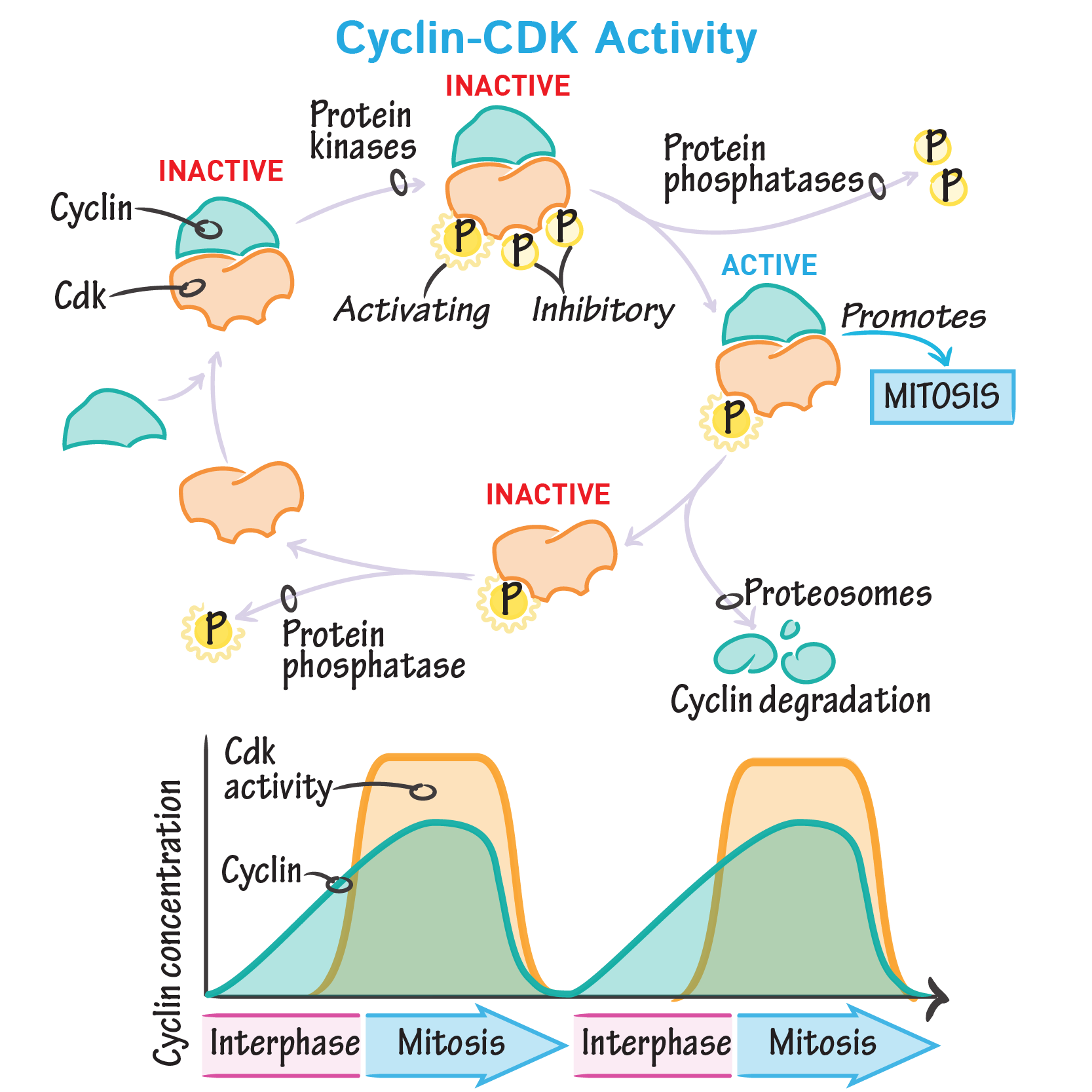
Cell growth can be referred to as cell division. Cell division is an active process that requires activation and inhibition of cell cycle proteins. Growth regulates the transition of the cell through two important checkpoints they are,
G0 ———–> G1
G1 ———–> S
These checkpoints are called DNA damage checkpoints, cells will not cross this checkpoint or in other words, cells are arrested if any DNA damage is found, in case of DNA damage, the genome is repaired by the cell and then proceeds to cell division. The cell cycle will not continue in the presence of active inhibition and the absence of stimulation. Growth factors provide active stimulation. The transition from G0 to G1 is controlled by growth factors in the following cells.
A skin cell, epidermal growth factor is the growth factor that regulates transition.
In connective tissue, the transition of such cells is regulated by fibroblast growth factor .
Mesenchymal cells are also regulated by fibroblast growth factor.
Nerve cells are regulated by the nerve growth factor .
Thrombus forming cell regulation of types of cell are controlled by the platelet derived growth factor .
Vascular Endothelial Growth Factor promotes angiogenesis, the process of formation of the blood vessels.
Vascular Endothelial Growths Factor And Receptors
a. Vascular endothelial growth factors
Vascular endothelial growth factors, also known as vascular permeability factor , or VEGF, constitute a sub-family of growth factors produced by cells that stimulate the formation of blood vessels and a mitogen for vascular endothelial cells. VEGFs are important signaling proteins involved in both vasculogenesis, the de novo formation of the embryonic circulatory system, and angiogenesis, the growth of blood vessels from pre-existing vasculature. There are five members in the vascular growth factor family, include VEGF-A, VEGF-B, VEGF-C, VEGF-D and placenta growth factor . VEGF-A is the first discovery member of the vascular endothelial growth factor family. Vascular endothelial growth factors are key mediators of angiogenesis that is crucial for development and metastasis of tumors. Overexpression of VEGF can contribute to disease. Solid cancers cannot grow beyond a limited size without an adequate blood supply cancers that can express VEGF are able to grow and metastasize. When VEGF is overexpressed, it can cause vascular disease in the retina of the eye and other parts of the body. Drugs can inhibit VEGF and control or slow those diseases, such as aflibercept, bevacizumab, ranibizumab and pegaptanib sodium .
b. Vascular endothelial growth factor Receptors
You May Like: Geometry Dash Toe 2
C Differentiation And Transformation
Cell differentiation has been shown to play an important role in the regulation of VEGF gene expression . The VEGF mRNA is up-regulated during the conversion of 3T3 preadipocytes into adipocytes or during the myogenic differentiation of C2C12 cells. Conversely, VEGF gene expression is repressed during the differentiation of the pheochromocytoma cell line PC12 into nonmalignant, neuron-like cells. These studies also indicate that induction of VEGF mRNA expression in preadipocytes requires pathways mediated by both protein kinase C and protein kinase A activation . Consistent with the presence of AP-1 and AP-2 sites in the VEGF gene promoter, phorbol esters and forskolin, a potent activator of adenylate cyclase, induce VEGF mRNA expression . Accordingly, luteotrophic hormone, a known activator of adenylate cyclase, has been shown to induce expression of VEGF mRNA in cultured bovine ovarian granulosa cells .
Taken together, these findings indicate that several, unrelated, alterations in cellular regulatory pathways result in VEGF up-regulation. Therefore, this event may be a final common pathway necessary for uncontrolled proliferation in vivo.
Engineering Gfs To Control Spatial And Temporal Presentation
Biomaterial-based delivery is a common strategy to efficiently deliver GFs. Immobilizing GFs within a biomaterial gives the possibility to achieve a sustained release and a localized delivery. Such approaches may considerably reduce the need for multiple doses and potentially reduce adverse effects. Therefore, various methods have been explored to enhance interactions between GFs and biomaterials.
Figure 1. GF engineering strategies in regenerative medicine. Strategies to control spatial and temporal presentations , stability , and signaling .
You May Like: Geometry Dash 1.9 Release Date
Epidermal Growth Factor Promotes Cell Division In The Absence Of This Growth Factor Is A Cell Likely To Continually Go Through All The Cell Cycle Phases
a. yes
A normal cell would is not permitted to pass to the next cycle if it is not “told” to do so by a growth factor. I don’t know how much detail you need, but growth factor receptor transduction pathways, so-called MAPK pathways culminate in hyperphosphorylation of retinoblastoma protein which acts as the main gate keeper to cycle progression. Hyperphosphoyrlated Rb leads to an increase in the G1/S cyclin which –and I’m skipping some details — leads to transition to S phase. Without growth factor signaling, a normal cell is unable to progress through those steps, and thus, remains in G1.