Mitochondrial Cristae Are Homologous To Alphaproteobacterial Icms
Given the alphaproteobacterial origin of MICOS and its critical function in the development of mitochondrial cristae, it is possible that cristae also have a pre-endosymbiotic origin. This possibility attains particular significance with the observation that several members of the Alphaproteobacteria develop intracytoplasmic membranes that resemble some crista morphotypes . The discovery of MICOS and its alphaproteobacterial roots now allows testing the hypothesis that mitochondrial cristae and alphaproteobacterial ICMs are homologous structures. Although direct experimental evidence for the involvement of alphaMic60 in ICM development is lacking, four main lines of evidence indicate that there has been a functional conservation in the evolution of eukaryotic Mic60 from alphaMic60. These include: the presence of a mitofilin signature domain in alphaMic60 , the structural conservation between alphaMic60 and eukaryotic Mic60 , the expression profile of alphaMic60 which reveals that alphaMic60 is overexpressed under conditions that promote ICM development in Rhodobacter sphaeroides , and the localization of alphaMic60 to ICMs, as revealed by proteomic studies of isolated ICMs of Rhodobacter sphaeroides , Rhodospirillum rubrum , and Rhodopseudomonas palustris . These observations strongly suggest that MICOS performs the same general function in both mitochondria and alphaproteobacteria and that there has been a historical continuity between cristae and ICMs .
Isolation Of Mitochondria And In Vitro Import
Mitochondria from mouse liver and HeLa cells were isolated, and mitochondrial import reactions were performed as described previously . Swollen mitochondria were obtained by incubation in 5 mM HEPES, pH 7.5, for 10 min on ice. Protease treatment was done by incubating mitochondria with 0.125 mg/ml trypsin for 20 min on ice and inactivated with soybean trypsin inhibitor . Mitochondrial lysates were size fractionated by SDS-PAGE and subject to autoradiography.
Why Can This Organism Survive Without Mitochondria
Without mitochondria , higher animals would likely not exist because their cells would only be able to obtain energy from anaerobic respiration , a process much less efficient than aerobic respiration.
What is the purpose of cristae membrane?
Which best describes the function of cristae?
Which best describes the function of cristae? They increase the surface area for reactions associated with cellular respiration.
Read Also: What Is Irb In Psychology
What Is Cristae Biology Class 9
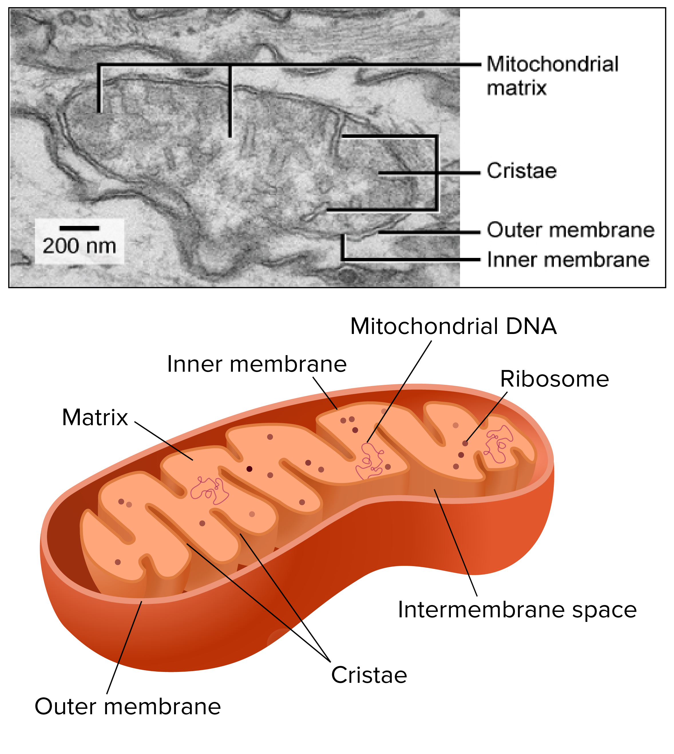
Cristae is the compartment in the inner mitochondrial membrane that expands the surface area of the inner mitochondrial membrane, enhancing its ability to produce ATP. Cristae are studded with F1 particles or oxysomes. Cristae are invaginations of the inner membrane that perform the chemiosmotic function.
Mitochondrial Fusion/fission Dynamics Contributes To The Formation Of Lamellar Cristae

In order to obtain more insight into the role of Mgm1 in the formation of cristae, we analyzed the biochemical composition and ultrastructure of mitochondria in the dnm1mgm1 double deletion mutant. Growth of the dnm1 and dnm1mgm1 strains on non-fermentable carbon sources was similar . On fermentable carbon source, however, the double deletion mutant displayed a high rate of loss of mtDNA, pointing to a residual function of Mgm1 in mitochondria of the dnm1 mutant . The steady state levels of a number of mitochondrial proteins were similar in WT, dnm1, and dnm1mgm1 strains . The interior of dnm1mgm1 mitochondria, like that of the dnm1 mutant, was full of tubular cristae. Strikingly, lamellar cristae were not observed in mitochondria of the dnm1mgm1 mutant . Thus, Mgm1 appears to be essential for the formation of lamellar cristae, but is not required for the formation of tubular cristae and CJs.
Growth, structure and protein composition of cristae in mutants lacking either Mgm1, Mic10, Mic60 or Su e in the Dnm1 deletion background.
3D reconstruction with modelling of a mitochondrion in the dnm1mgm1 double deletion mutant.
Green, IBM and cristae connected to the IBM blue, tubular cristae without visible connection to the IBM yellow, tubular cristae without connection to the IBM.
3D reconstruction with modelling of a mitochondrion in the dnm1mic60 double deletion mutant.
Don’t Miss: How To Do Unit Conversions In Chemistry
Cell Culture Rna Interference Transfection And Retroviral Transductions
Transfections of expression constructs were performed with FuGENE 6 reagent . Mitotracker Red CMXRos was used to stain mitochondria as per manufacturer’s protocol. A Nikon confocal microscope was used to scan the transfected cells. The images were obtained in the EZ-C1 acquisition software with a 60Ã objective lens and 0.75 numerical aperture. The images were acquired at room temperature in Slowfade by using a Nikon D-eclipse C1 camera.
RNAi experiments were performed in HeLa cells essentially as described previously . The complementary RNA oligonucleotides AAUUGCUGGAGCUGGCCUUTT and AAGGCCAGCUCCAGCAAUUTT were derived from nucleotides 135â155 of the mitofilin cDNA. A pair of nonspecific scramble RNA oligonucleotides was used as a control . The short hairpin RNAi construct was generated by PCR with the primers 5â² TGCTCTAGAAAAAAAGCTACCTGAAGTAGAATATCTACGGCACGCAAGCTTCCATGCCGCAGATACTCTACTTCAGGCAGCGGTGTTTCGTCCTTTCCACAA 3â² and 5â² CGCGGATCCAAGGTCGGGCAGGAAGAGGGC 3â² and cloning downstream of the human U6 promoter into the XbaI and BamHI sites of pAVU6 vector. Two rounds of transfections were performed and monitored using Su9-GFP and Su9-RFP for the first and second rounds, respectively. The reporter constructs were used at 1:10 ratio relative to the mitofilin expression and RNAi vectors.
What Happens In The Cristae
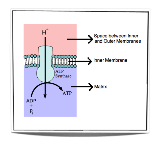
The mitochondrial cristae are where electrons are passed through the electron transport chain, which pumps protons to power the production of energy molecules called ATP. All of this results in the pumping of hydrogen ions, the conversion of oxygen gas into water, and the production of ATP.
Is ATP made in the cristae?
The electron transport chain and chemiosmosis takes place on this membrane as part of cellular respiration to create ATP and can be seen in the diagram: The cristae increase the surface area of the inner membrane, allowing for faster production of ATP because there are more places to perform the process.
What do the cristae do within the mitochondria?
Mitochondrial cristae are the folds within the inner mitochondrial membrane. These folds allow for increased surface area in which chemical reactions, such as the redox reactions, can take place.
What are Cristae short answer?
Cristae is the compartment in the inner mitochondrial membrane that expands the surface area of the inner mitochondrial membrane, enhancing its ability to produce ATP. Cristae are studded with F1 particles or oxysomes. Cristae are invaginations of the inner membrane that perform the chemiosmotic function.
Also Check: Why Study Geography At University
Blue Native Gel Electrophoresis
75 g of mitochondria were pelleted by centrifugation and resuspended in BN-lysis buffer . For analysis of F1FO-ATP synthase or MICOS complex 1 % or 3 % digitonin were used. Cleared lysates were supplemented with Native PAGE 5% G-250 Sample Additive and subjected to BN-PAGE . After blotting on PVDF membranes immuno-decoration using the indicated antibodies was performed.
Mitochondria Lacking Dnm1 Show Altered Crista Structure
Structure and protein composition of cristae in cells deficient in Dnm1.
Ultrastructure and quantitative evaluation of mitochondria in dnm1 cells grown on YPG . Tomographic reconstruction of a mitochondrion of a dnm1 cell. Green, IBM and cristae connected to the IBM blue, tubular cristae without visible connection to the IBM. Steady state levels of mitochondrial proteins of dnm1 cells. WT and dnm1 cells were grown on YPG, aliquots of mitochondrial protein were analyzed by SDS-PAGE and immunoblotting. Assembly state of MICOS and of F1FO in dnm1 cells. Analysis as in Figure 4D. Arrow head, assembled MICOS complex.
3D reconstruction with modelling of a mitochondrion in the dnm1 mutant.
Green, IBM and cristae connected to the IBM blue, tubular cristae without visible connection to the IBM yellow, tubular cristae without connection to the IBM.
Recommended Reading: How To Solve Oxidation Number In Chemistry
Atp Synthase Forms Rows Of Dimers In Crista Membranes
The mitochondrial F1-Fo ATP synthase is the most conspicuous protein complex in the cristae. The ATP synthase is an ancient nanomachine that uses the electrochemical proton gradient across the inner mitochondrial membrane to produce ATP by rotatory catalysis . Protons moving through the Fo complex in the membrane drive a rotor ring composed of 8 or 10 c-subunits. The central stalk propagates the c-ring torque to the catalytic F1 head, where ATP is generated from ADP and phosphate through a sequence of conformational changes. The peripheral stalk prevents unproductive rotation of the F1 head against the Fo complex.
For many years it was assumed that the ATP synthase and other energy-converting complexes are randomly distributed over the inner membrane. The first hint that this is not the case came from deep-etch freeze-fracture electron microscopy, which revealed double rows of macromolecular complexes in the tubular cristae of the single-cell ciliate Paramecium . The double rows were thought to be linear arrays of mitochondrial ATP synthase. This is indeed what they are, but it could only be shown unambiguously more than 20 years later by cryo-ET , which revealed rows of ATP synthase dimers in mitochondria of all species investigated . Until then, the rows were thought to be peculiar to Paramecium.
Fig. 4
Where Is Cisternae Found
The Golgi apparatus The Golgi apparatus, also called Golgi complex or Golgi body, is a membrane-bound organelle found in eukaryotic cells that is made up of a series of flattened stacked pouches called cisternae. It is located in the cytoplasm next to the endoplasmic reticulum and near the cell nucleus.
Also Check: What Are Some Examples Of Human Geography
What Is The Structure And Function Of The Extracellular Matrix
The extracellular matrix is a structural support network made up of diverse proteins, sugars and other components. It influences a wide number of cellular processes including migration, wound healing and differentiation, all of which is of particular interest to researchers in the field of tissue engineering.
What Are Cristae Short Answer

Cristae is the compartment in the inner mitochondrial membrane that expands the surface area of the inner mitochondrial membrane, enhancing its ability to produce ATP. Cristae are studded with F1 particles or oxysomes. Cristae are invaginations of the inner membrane that perform the chemiosmotic function.
Don’t Miss: What Is The Physics Behind Roller Coasters
What Is The Difference Between Cristae And Cisternae
Cristae are found in mitochondria and are a fold in their inner membrane while cisternae are found in the Endoplasmic reticulum and Golgi apparatus in the form of flattened membrane discs. Cristae have proteins, including ATP synthase and many cytochromes while cisternae have several enzymes active inside them.
The Micos Complex At Center Stage Of Cristae Remodeling
J. Cell Biol.EMBO J.
J. Cell Biol.Dev. Cell.
J. Cell Biol.J. Cell Biol.Biol. Chem.J. Intern. Med.Biochim. Biophys. Acta.Biol. Chem.J. Intern. Med.
Biochim. BiophysActa.Int. J. Mol. Sci.Biol. Chem.J. Biochem.Biochim. Biophys. Acta.
J. Med. Genet.
J. Cell Biol.Mol. Biol. Cell.
J. Cell Biol.
J. Cell Biol.
Paracoccus denitrificansEscherichia coli
J. Cell Biol.Curr. Biol.Curr. Biol.
J. Cell Biol.Cell Metab.Nat. Commun.Nat. Commun.
in vitro
J. Cell Biol.
Cell Metab.
de novoEMBO J.
- Munoz-Gomez S.A.
Curr. Biol.
Biochim. Biophys. Acta.PLoS One.Elife.J. Mol. Biol.
You May Like: What Jobs Require Biology Degree
What Do The Cristae In The Mitochondria Contain
The inner membrane forms invaginations, called cristae, that extend deeply into the matrix. The crista lumen contains large amounts of the small soluble electron carrier protein cytochrome c. The mitochondrial cristae are thus the main site of biological energy conversion in all non-photosynthetic eukaryotes.
What Are Two Functions Of The Inner Membrane
Biological membranes have three primary functions: they keep toxic substances out of the cell they contain receptors and channels that allow specific molecules, such as ions, nutrients, wastes, and metabolic products, that mediate cellular and extracellular activities to pass between organelles and between the
Don’t Miss: What Kind Of Math Is Needed For Nursing
Mitofilin Is Localized To A Subcompartment Within The Intermembrane Space
Figure 1. Characterization of the putative mitochondrial targeting sequence and membrane-anchoring domain of mitofilin. Mitofilin, mitofilin -DHFR, and Su9-DHFR were synthesized in rabbit reticulocyte lysate as the precursor form in the presence of methionine and imported into isolated mouse liver mitochondria. After import, the mitochondria were untreated , treated directly with trypsin , or treated with trypsin in the presence of hypoosmotic shock. Soybean trypsin inhibitor also was included in the lane 6. The mitochondria were recovered through centrifugation , whereas the supernatant contained untargeted protein. Alkaline extraction was performed by resuspending the mitochondrial pellets in 100 mM Na2CO3 for 30 min on ice. The mitochondrial membranes were recovered by ultracentrifugation at 100,000 Ã g. All the samples were size-fractionated by SDS-PAGE and subjected to autoradiography. The precursor and mature forms of the proteins are indicated as p and m, respectively.
Figure 2. Intramitochondrial localization of mitofilin. Cos7 cells expressing fusion proteins mitofilin-GFP, mitofilin -GFP, Tim23-GFP and HeLa cells stably expressing cytochrome c-GFP were stained with Mitotracker . The localizations of the GFP fusion proteins and Mitotracker were assessed by confocal microscopy. Bar, 10 μm.
Mitochondrial Inner Membrane Topology
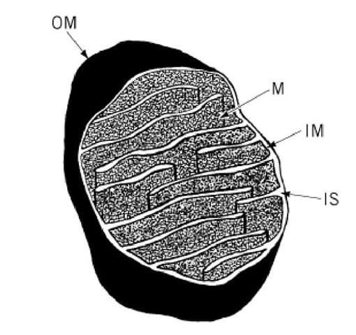
The early model of mitochondrial structure portrayed the cristae as baffle-like folds in the inner membrane . Three-dimensional images provided by electron tomography of a wide variety of mitochondria, isolated and in situ, indicate a different internal architecture. The inner membrane comprises numerous pleiomorphic compartments, which are connected to each other and to the periphery of the inner membrane by narrow tubular segments . These tubular connecting regions can vary in length from a few tens to several hundred nanometers.
Functional Implications of the Cristae Junctions
The fact that cristae open into the intermembrane space through narrow, sometimes very long tubular segments suggests that diffusion of solutes between the internal compartments of mitochondria might be restricted. This was not a concern with the early structural model, which depicted the baffle-like cristae with wide, slot-shaped openings into the intermembrane compartment. Computer simulations indicate that diffusion of ADP between the intermembrane and intracristal compartments via long, narrow tubes can cause depletion of ADP in the intracristal space and locally diminished ADP phosphorylation. Thus, the topology of the mitochondrial inner membrane may directly influence the efficiency of ATP generation by the organelle.
Cristae Junctions Form Spontaneously
Guy D. Potter M.D., in, 1971
Don’t Miss: What Is Pcr In Biology
Formation Of Cristae Creating More Mitochondrial Inner Membrane Surface Area And Protonic Capacitance
From the protonic membrane capacitor of Fig. , it is quite apparent that mitochondrial cristae formation creates more mitochondrial inner membrane surface area ), which immediately contributes to its membrane capacitance ). Therefore, its bioenergetics significance can now be better understood with the Lee model,,,, of transmembrane electrostatically localized protons as shown in the membrane potential equation , which shows that exists precisely because of the excess cations and the excess anions charge layers localized on the two sides of the membrane in a protons-membrane-anions capacitor structure . This also explains the origin of and its relationship with the concentration of electrostatically localized protons \ as expressed in Eqs. and .
Figure 3
Comparing the numbers of transmembrane electrostatically localized protons per mitochondrion for a mitochondrion without cristae and with cristae as a function of membrane potential .
What Is The Cristae Quizlet

Cristae. Finger -like projections that provide a large surface area for reactions of cellular respiration. Matrix. Space inside a mitochondrion. Glycolysis.
What is the importance of cristae in mitochondria?
To increase the capacity of the mitochondrion to synthesize ATP, the inner membrane is folded to form cristae. These folds allow a much greater amount of electron transport chain enzymes and ATP synthase to be packed into the mitochondrion.
What happens at the cristae?
The mitochondrial cristae are where electrons are passed through the electron transport chain, which pumps protons to power the production of energy molecules called ATP. All of this results in the pumping of hydrogen ions, the conversion of oxygen gas into water, and the production of ATP.
You May Like: How Did The Geography Of Greece Affect Its Development
What Is Function Golgi Apparatus
The Golgi apparatus, or Golgi complex, functions as a factory in which proteins received from the ER are further processed and sorted for transport to their eventual destinations: lysosomes, the plasma membrane, or secretion. In addition, as noted earlier, glycolipids and sphingomyelin are synthesized within the Golgi.
Strains And Growth Conditions
The strains and plasmids used are listed in Tables S2 and S3. Culturing of yeast strains was performed using standard methods at 30°C on complete liquid media containing 2% lactate. Strains containing plasmids were grown on selective liquid media containing 2% lactate supplemented with 0.1% glucose. For overexpression of Fcj1, the wild type and the fcj1 strain containing the plasmid pCM189-Fcj1 were grown in the absence of doxycycline. For down-regulation of Fcj1, the same plasmid was used in a fcj1 strain, and 20 µg/ml doxycycline was added to the medium. Drop dilution growth tests were performed with 1:10 dilution steps and incubation on yeast peptone dextrose and lactate medium plates for 24 d at 24°C. Rho0/rho cell formation was determined on complete glycerol plates supplemented with 0.1% glucose.
Don’t Miss: What Math Class Do 11th Graders Take
What Is Cristae And Matrix
Each membrane is a phospholipid bilayer embedded with proteins. The inner layer has folds called cristae, which increase the surface area of the inner membrane. The area surrounded by the folds is called the mitochondrial matrix. The cristae and the matrix have different roles in cellular respiration.
Membrane Rearrangement During Cellular Ageing
Ageing is a fundamental yet poorly understood biological process that affects all eukaryotic life. Deterioration in mitochondria is clearly seen in ageing, but details of the underlying molecular events are largely unknown. Cryo-ET of mitochondria from the short-lived model organism Podospora anserina revealed profound age-dependent changes in the inner membrane architecture . In normal mitochondria of young cells, the cristae protrude deeply into the matrix. Formation of cristae depends both on the rows of ATP synthase dimers along the edges and on the MICOS complex at the crista junctions . With increasing age, the cristae recede into the inner boundary membrane and the inter-membrane space widens. The MICOS complex most likely has to come apart for this to happen. Eventually, the matrix breaks up into spherical vesicles within the outer membrane. The ATP synthase dimer rows disperse and the dimers dissociate into monomers. As the inner membrane vesiculates, the sharp local curvature at the dimer rows inverts, so that the ATP synthase monomers are surrounded by a shallow concave membrane environment, rather than the sharply convex curvature at the crista ridges . Finally, the outer membrane ruptures, releasing the inner membrane vesicles, along with apoptogenic cytochrome c, into the cytoplasm. Cytochrome c activates a cascade of proteolytic caspases, which degrade cellular proteins . The cell enters into apoptosis and dies.
Fig. 9
Read Also: What Is Reference Point In Physics