Dubs Involved In Ciliogenesis During G0/g1
Many DUBs have been found to be required for the formation of primary cilia during G0/G1 phase of the cell cycle, a process termed ciliogenesis. Firstly, the DUB CYLD is recruited to centrosomes and the basal body of cilia via its interaction with CAP350 , where it has to be present and catalytically active to promote docking of basal bodies at the plasma membrane and hence ciliogenesis . A concurrent study also demonstrated that CYLD is required for docking of basal bodies at the plasma membrane and identified that this can, at least in part, be explained by its ability to deubiquitylate CEP70 . Deubiquitylation of CEP70 allows it to interact with -tubulin at the centrosome to mediate ciliogenesis . In addition, CYLD inactivates HDAC6, which modulates cilia length . Secondly, via an independent mechanism to its roles in centrosome duplication, USP9X also regulates ciliogenesis . During G0/G1, USP9X is recruited to the centrosome where it deubiquitylates and stabilises NPHP5 , a positive regulator of ciliogenesis, so favouring cilia formation. However, at G2/M, USP9X becomes cytoplasmic, allowing degradation of NPHP5 and loss of cilia. Finally, a survey of DUB subcellular localisation found that USP21 localised to centrosomes and microtubules . USP21 is required for effective microtubule regrowth from centrosomes, neurite outgrowth, generation of the primary cilium and hedgehog signalling .
Dubs Affecting Centrosome Maturation Separation And Mitotic Spindle Organisation During G2 And Mitosis
BRCA1 /BARD1 -dependent ubiquitylation of -tubulin plays a key role in the regulation of centrosome duplication and microtubule nucleation, with BRCA1 loss resulting in centrosome amplification . An siRNA screen for DUBs that affect levels of ubiquitylated -tubulin identified BAP1 and UCHL1 as candidates . While UCHL1 interacts with -tubulin in G1, the BAP1 interaction is largely confined to mitosis, suggesting that these two DUBs regulate -tubulin in a cell cycle-dependent manner . BAP1 removes ubiquitin from -tubulin, and mitotic defects in cells with low BAP1 levels are rescued by expression of BAP1 but not a catalytically inactive mutant. While the mechanism remains to be fully elucidated, it seems that deubiquitylation of -tubulin by BAP1 during mitosis allows proper spindle organisation and function . CEP192 is a centrosomal protein with roles in maturation of centrosomes at the onset of mitosis and organisation of the mitotic microtubule landscape. Mass spectrometry identified the deubiquitylase CYLD as a CEP192 interactor, and CYLD co-depletion restores spindle assembly defects in CEP192-depleted cells .
Are Centrosomes Essential For All Animal Cells
Maybe not. Although centrosome has a key role in efficient mitosis in animal cells, the centrosome is not essential for mitosis in certain animal species like fly and flatworm. In an experiment done in the fruit fly , even the centrioles were destroyed by a laser, the mitosis proceeds normally with a morphologically normal spindle. Moreover, the fruit fly grows normally. This suggests the cells should have other ways to organize their microtubules.
Recommended Reading: What Is Prediction In Geography
Are Centrosomes Ever Important For Cell Division
Centrosomes are important for specialized cell divisions. For example, in Drosophila, adult males with no centrosomes show highly abnormal meiotic divisions . Moreover, eggs from mothers that are mutant for centriole proteins arrest very early in embryonic development after only a few abnormal mitoses, showing that centrioles are necessary for syncytial mitoses . Moreover, asymmetric cell divisions can also be abnormal in the absence of centrosomes . In summary, whereas centrioles may be dispensable for cell division in some tissues of the fly, they are absolutely essential in others, perhaps due to tissue specificity constraints, such as weaker checkpoints, different cell size and/or sharing of common cytoplasm in the context of a syncytium. The same is true in other organisms, such as the Caenorhabditis elegans embryo and fission yeast, where the centrosome and its equivalent, the spindle pole body, are essential for bipolar spindle assembly and cytokinesis, respectively .
Mechanisms Controlling Centrosome Positioning In Immune Cells

In summary, centrosomes in immune cells contribute to the immune response through polarized delivery of lytic granules and cytokines, a process that is accompanied by centrosome translocation. Less is known about the detailed mechanisms controlling centrosome behavior though actin and microtubule dynamics, which generate forces that pull the centrosome toward the IS. Furthermore, the signals that drive centrosome movement and thus initiate or terminate the immune response also warrant further exploration.
Also Check: Answers To Odysseyware Algebra 1
Similarities Between The Centrioles Cilium And Flagellum:
- Centriole, cilium and flagellum resemble one another in their broad structure and function.
- All of them are made up of microtubules.
- The three possess nine peripheral fibrils of microtubules. Fibril organization is 9 + 0 in centrioles and 9 + 2 in case of cilia and flagella.
- Basal granule present at the base of a cilium or flagellum is derived from a centriole and resembles the same in structure.
- All the three are capable of movements. Centriole does so to a limited extent inside the cytoplasm. A cilium or flagellum produces a current in an external liquid medium for locomotion, feeding, aeration and circulation.
- Centrioles are parent organelles which produce basal bodies, cilia and flagella. They have nucleating centres or massules for the growth of microtubules.
What Is A Centrosome
I know that a centrosme is composed of two perpendicular centrioles, but the following sentences of Wikipedia confuse me:
Interestingly, centrioles are not required for the progression of mitosis.
Many cells can completely undergo interphase without centrioles.
Unlike centrioles, centrosomes are required for survival of the organism.
If centrosomes are essential then does this doesn’t imply that centrioles too are necessary since centrosome is made of two centrioles, if not then what is it made of?
Reading the specified Wikipedia article about Centrosome, actually explains why centrioles are NOT IMPORTANT for the PROGRESSION OF MITOSIS.
To better understand what wiki meant, let us look at both centrioles& centrosomes.
According to Biology Pages for Centrioles and Centrosomes > >
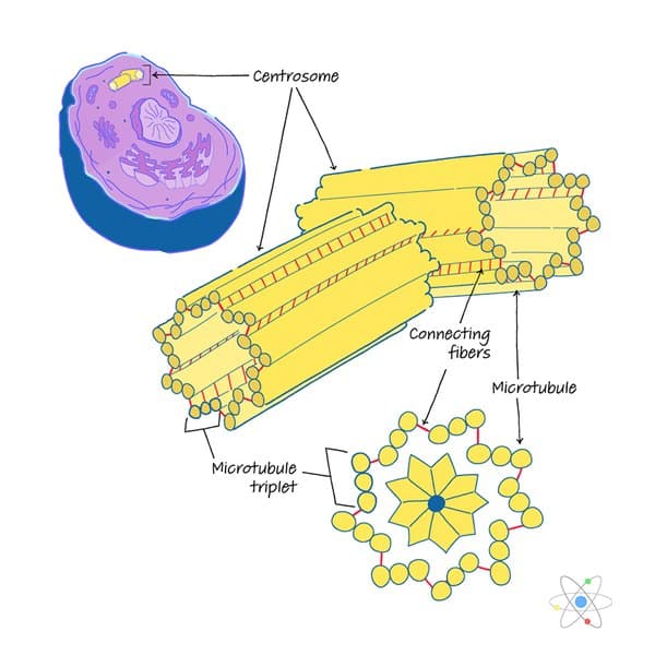
- Centrioles are built from a cylindrical array of 9 microtubules, each of which has attached to 2 partial microtubules.
- is located in the cytoplasm usually close to the nucleus.
- It consists of two centrioles oriented at right angles to eachother embedded in a mass of amorphous material containing more than100 different proteins.
- It is duplicated during S phase of the cell cycle.
- Just before mitosis, the two centrosomes move apart until theyare on opposite sides of the nucleus.
- As mitosis proceeds, microtubules grow out from eachcentrosome with their plus ends growing toward the metaphaseplate. These clusters of microtubules are called spindlefibers.
Read Also: What Is Nuclear Fusion Chemistry
Dubs With Roles In Mitotic Progression And Cytokinesis
Following replication of the genome, and assuming checkpoints are satisfied in G2, the cell enters mitosis, where the newly replicated sister chromatids must be divided into each daughter cell. To achieve this, the cell passes through a sequence of distinct mitotic phases: prophase, metaphase, anaphase, telophase and cytokinesis . Prior to mitosis, the mitotic kinase CDK1 is held in an inactivate state by WEE1 phosphorylation, until SCFTrCP-mediated ubiquitylation and degradation of WEE1 triggers mitotic entry USP50 can repress mitotic entry through stabilising WEE1 . Subsequently, USP7 can indirectly regulate the levels of Aurora A, a kinase required for correct maturation of the bipolar mitotic spindle, by stabilising CHFR , an E3 ligase that targets Aurora A for degradation .
The DUB CYLD plays roles during both metaphase and cytokinesis. CYLD directly interacts with the catalytic domain of HDAC6 , inhibiting -tubulin deacetylation and therefore indirectly increasing the stability of microtubules. This governance of microtubule stability by CYLD plays a role in spindle orientation during metaphase and regulates the rate of cytokinesis . Finally, USP8 and AMSH , two DUBs that are usually recruited to endosomes, have an important role in cytokinesis. The scission of the two daughter cells requires components of the ESCRT machinery including VAMP8 , which co-localises with, and is deubiquitylated by, both USP8 and AMSH during cytokinesis .
Dubs Associated With The Centrosome Cycle
Many human cells also display cilia in a cell cycle-dependent manner. During G1 centrosomes migrate to the cell cortex, where the mother centriole matures into a basal body which acts as a template for cilia elongation. During S-phase, both mother and daughter centrioles undergo duplication as normal. Then, prior to mitosis, cilia disassemble and the centrioles migrate back to the cell interior, ready to act as spindle poles during mitosis .
Also Check: What To Expect In Physics Class
Breaking The Ties That Bind: New Advances In Centrosome Biology
- Abbreviations used in this paper:
J Cell Biol
Balca R. Mardin, Elmar Schiebel Breaking the ties that bind: New advances in centrosome biology. J Cell Biol 2 April 2012 197 : 1118. doi:
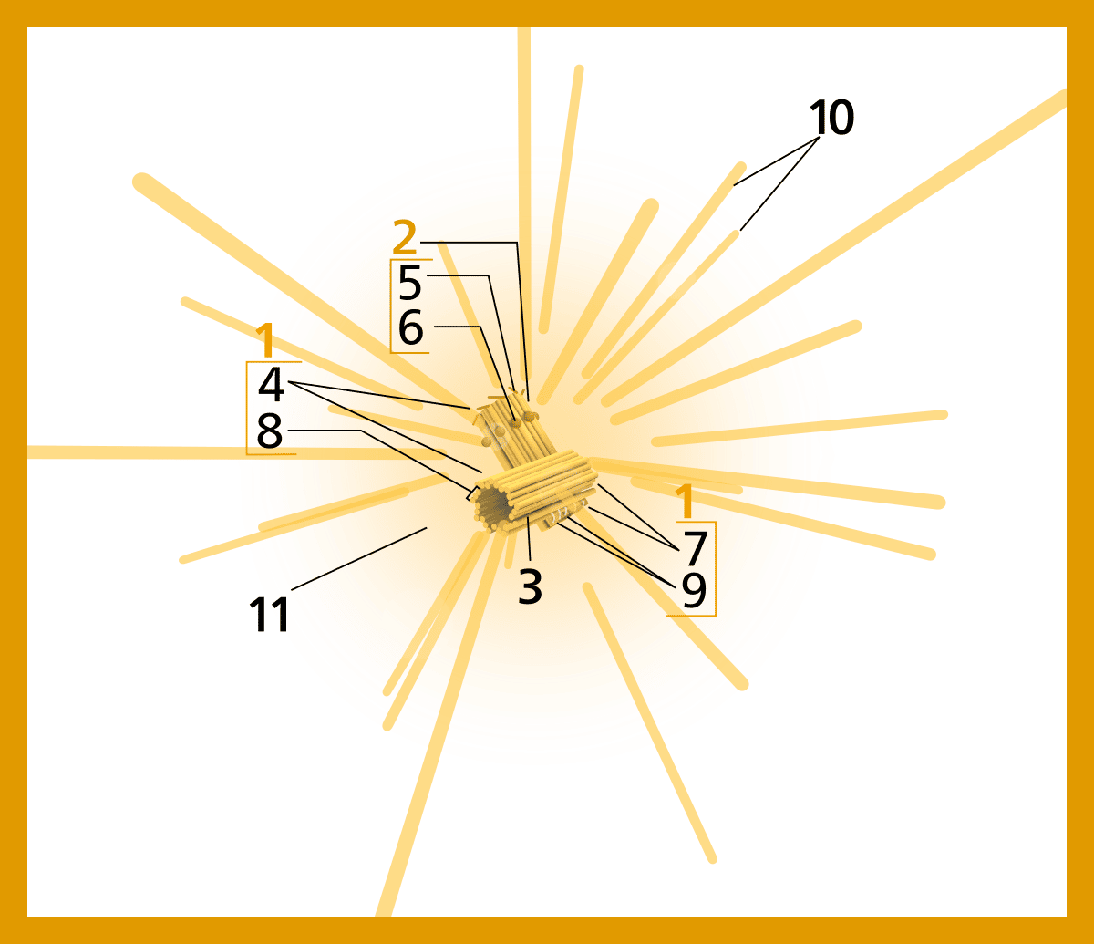
The centrosome, which consists of two centrioles and the surrounding pericentriolar material, is the primary microtubule-organizing center in animal cells. Like chromosomes, centrosomes duplicate once per cell cycle and defects that lead to abnormalities in the number of centrosomes result in genomic instability, a hallmark of most cancer cells. Increasing evidence suggests that the separation of the two centrioles is required for centrosome duplication. After centriole disengagement, a proteinaceous linker is established that still connects the two centrioles. In G2, this linker is resolved , thereby allowing the centrosomes to separate and form the poles of the bipolar spindle. Recent work has identified new players that regulate these two processes and revealed unexpected mechanisms controlling the centrosome cycle.
Centrosome In Animal Cells
In most animal cells, centrosomes are not required in the cell division process even though they add to the effectiveness of the mitotic spindle arrangement. In humans, dysfunctioning of centrosomes can stimulate cancer as a result of an increase in the levels of instability in chromosomes or due to the metastatic capability of cancer cells. However, the study on this lacks evidence.
You May Like: Honors Algebra 2 Linear Function Word Problems Answers
What About The Pericentriolar Material How Do Centrioles Recruit It And What Does It Do
Several centrosome components have been identified recently, through proteomic studies or functional genomic analysis and their localization and function characterized . These include CEP192/SPD2 , CEP152/asl , Pericentrin, SAS4/CPAP and CNN /CDK5RAP2 , which bind to centrioles and/or to each other and recruit microtubule nucleators, such as gamma-tubulin. From these studies, a new view, of a highly organized PCM is emerging, where different domains might be involved in separate functions and are regulated differently through the cell cycle . The size and organization of the PCM is likely to impinge on centrosome function and is determined by the intrinsic properties of its components , their availability and their regulation by kinases . How this all works to ensure centrosome function is poorly understood and is an important avenue of research for the future.
Regulation Of The Cell Cycle And Centrosome Biology By Deubiquitylases

Correspondence:
Sarah Darling, Andrew B. Fielding, Dorota Sabat-Popiech, Ian A. Prior, Judy M. Coulson Regulation of the cell cycle and centrosome biology by deubiquitylases. Biochem Soc Trans 15 October 2017 45 : 11251136. doi:
Also Check: What Is The Hardest Level In Geometry Dash
Centrosome: Useful Notes On Centrosome
ADVERTISEMENTS:
This article provides information about Centrosome, its Structure, Chemical composition, Functions and Origin!
Centrosome or cell centre is the area of cytoplasm around the centriole.
It is also called micro-centrum. Centrosome is found lying in the centre of the cell, near the nucleus, in the cytoplasm. In Metazoa, centrosome lies outside nucleus, but in Protozoa it lies within the nucleus.
Image Courtesy : http://upload.wikimedia.org/wikipedia/commons/7/7a/Centrosome_Cycle.svg
ADVERTISEMENTS:
It is not a universal cell constituent as it is lacking in some plant cells. They were discovered by Van Benden , in cells of certain parasites of cephalopods, indicating it as origin of aster. Later on, T. Boveri in 1888 described it in detail. The substance of centrosome is called kinoplasm which consists of two parts
Smaller bodies or centrioles.
Surrounding mass or centrosphere.
How Is A Centrosome Different From A Centromere
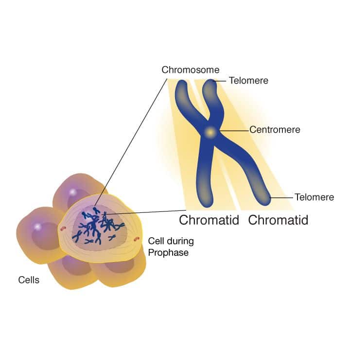
The centrosome is the organelle that contains two centrioles. Whereas centromere is a highly constricted region on the chromosome. A centrosome is a microtubule-organizing centre, whereas, centromere holds together the sister chromatids in a replicated chromosome.
Put your understanding of this concept to test by answering a few MCQs. Click Start Quiz to begin!
Select the correct answer and click on the Finish buttonCheck your score and answers at the end of the quiz
Recommended Reading: What Subjects Do You Need To Study Biological Sciences
Ii Organizing Microtubule Networks In The Cells
Centrosomes as a microtubule organizing center control the distribution of microtubule networks inside the cells. Microtubule is a versatile cytoskeleton, and the formation of the mitotic spindle is just one of its functions. In non-dividing cells, microtubules also serve as an intracellular highway to transport molecules and organelles within the cells. There is a group of motor proteins that can carry cargos while walking along the cytoskeleton, which is just like many small trucks driving on an intracellular transportation system.
The microtubule organization varies according to the cell types and cell cycle. It determines the internal organization of organelles and vesicular trafficking that benefit the cell functions.
Centrosomes can also orchestrate large changes to cell membrane shape under other circumstances, such as cell movement or phagocytosis.
Extended read:
Centrosomes In Plant Cells
Centrosomes in Plant Cells Plants and growths that don’t have centrosomes subsequently utilize MTOC structures to coordinate microtubules. Plant cells don’t have axle post bodies or centrioles with the exception of in flogging male gametes which are totally present in a couple of blooming plants.
The essential capacity of the MTOC for shaft association and microtubule nucleation gives off an impression of being taken up by the atomic envelope while mitosis of the plant cell.
Plants and fungi do not have centrosomes. Plant cells do not have spindle pole bodies or centrioles except in flagellated male gametes that are found in a few flowering plants only. The main function of MTOC for spindle organization and microtubule nucleation appears to be taken by the nuclear envelope during mitosis of the plant cells.
Animal and plant cells share the main cytoskeletal elements that help in controlled working. Plants do not have centrosomes resembling organelles but they are capable of building spindles and have developed cytoskeletal arrays such as preprophase band, the cortical arrays, and the phragmoplast that take part in the fundamental growth processes.
Also Check: What Are Physical Features In Geography
Word Games: Centrosome Vs Centrioles
Posted on 1/27/21 by Laura Snider
As is the case in A& P, keeping track of terminology can be one of the most challenging parts of a biology course, especially when youre studying cells. There are a lot of words that sound alike, and the structures they apply to may even have similar functions.
In this blog post, well be going over two particularly similar-sounding cellular structures: the centrosome and centrioles. To help you distinguish between them, well talk about the etymology behind each term, as well as the function of each structure.
Avoiding More Potential Confusion: Centromeres
Also, remember not to confuse either of these with the centromere . The word centromere shares the same Latin root but its Greek component, mere, refers to a partso, centromere literally means the center part of the chromosome.
The centromere of a chromosome. Screenshot from Visible Biology .
For more scientific vocabulary fun, check out these related VB Blog posts:
Read Also: College Algebra Sample Test Questions
What Is The Function Of The Centrosome
The centrosome has several functions. The central one is as the major microtubule organizing center in proliferating animal cells: thus, it helps to organize the microtubules that form the mitotic spindle in dividing cells, and orchestrate a wide variety of cellular processes, including cell motility, signaling, adhesion, coordination of protein trafficking by the microtubule cytoskeleton and the acquisition of polarity. The centrosome has crucial links to the nucleus, the Golgi, cell to cell junctions and acto-myosin cytoskeleton that are very important in positioning it and thus shaping the microtubule cytoskeleton in relation to the cell and the organism . The role of the centrosome in organizing cellular microtubules can differ from cell to cell and be regulated differently in different phases of the life of a cell.
How Does The Centrosome Perform Its Organizing Function

Although microtubules can form spontaneously from high concentrations of tubulin in vitro, in cells they are nucleated by specialized microtubule-nucleating proteins, some of which are associated with the centrosome. The centrosome is composed of two barrel-shaped microtubule-based organelles, the centrioles, surrounded by proteins collectively called the pericentriolar material . Proteins of both the centriole and the PCM can nucleate and anchor microtubules .
The role of the centrosome in directing cellular protein traffic depends upon the intrinsic polarity of microtubules, and microtubule-associated motor proteins that move differentially towards one microtubule end or the other. By nucleating microtubules, the centrosome thus determines the tracks along which different cellular components can be transported to different parts of the cell. It can also help to define the speed at which components move along those tracks, and act as a signaling center to modify some components before they are transported to their destinations.
You May Like: What Is Quantitative Data Psychology
Cell Biology Of Centrosome Asymmetry
Mother and daughter centrosomes/centrioles show different characteristics during the cell cycle and cell division. For instance, mother and daughter centrioles have different motility within the cell, where in G1 cells, the mother centriole is typically less motile than the daughter, presumably because of microtubules that the mother anchors.8 Piel et al. further demonstrated that mother centrioles move very close to the midbody, right before the abscission.9 Abscission, or the final stage of cytokinesis, occurs only from one side, where secretory vesicles carrying membrane/protein components required for abscission are carried only from the daughter centrosome-containing cell, leaving a midbody ring in the cell with the mother centrosome . As a result of such asymmetric inheritance of the midbody ring, some HeLa cells contain multiple remnant rings.10 Recently, Pohl et al. demonstrated that these ring remnants are typically digested by autophagy in normal cells, such that normal cells do not have multiple rings.11 As such, possession of multiple ring remnants might be a feature of abnormal cells. Elaborate mechanisms to remove midbody rings in normal cells suggest its importance in the maintenance of cellular homeostasis. It is also possible that the midbody ring is associated with unwanted cellular material, such as cell cycle regulators that drive cells into undesired cell cycle arrest or proliferation, or cell fate determinants .