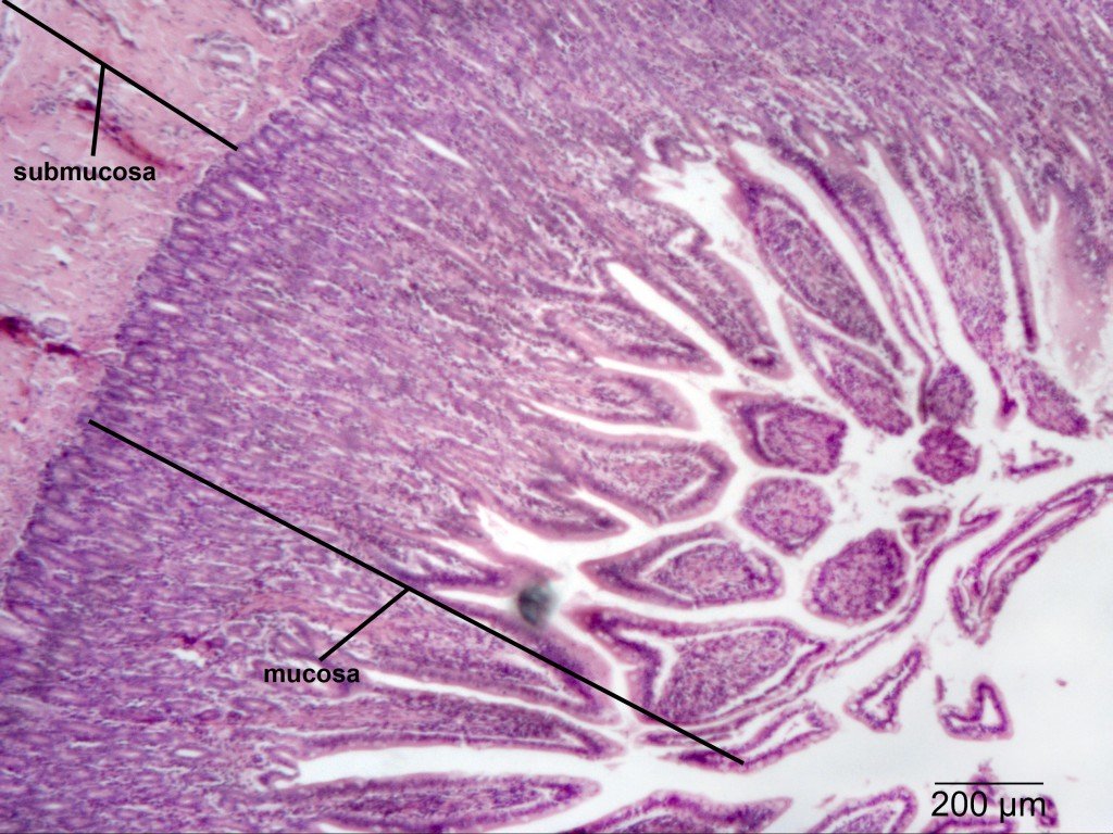
S Of The Small Intestine
The small intestine is further divided into three sections: the duodenum, the jejunum, and the ileum. The duodenum is the first and shortest section of the small intestine, which measures about fifteen inches long. It receives chyme from our stomachs. The duodenums intestinal cells also secrete amylase, sucrase, and lipase enzymes that break down fats and sugars.
The jejunum follows suit and is located near our belly buttons. The jejunum marks the end of our digestion of fats and carbohydrates. It is covered in villi and microvilli that make it the principal site of digestion. It is also a coiled structure that is thicker and has more blood vessels than the third and final section, the ileum. The ileum lies in our pelvic area, more or less, and is thinner and less vascular than the jejunum. The ileums main role is in absorption and it will absorb amino acids, lipids, fat-soluble vitamins, and vitamin B12.
Embryology And Anatomy Of The Duodenum
The duodenum develops during the fourth week of gestation from the distal foregut, proximal midgut, and the adjacent splanchnic mesenchyme. The junction of the foregut and midgut occurs in the second part of the duodenum, slightly distal to the major papilla. As the stomach rotates,so too does the duodenum, therefore developing a C-shaped configuration. During weeks 5 and 6 of embryologic development, the duodenal lumen is temporarily obliterated owing to proliferation of its mucosal lining. During the following weeks, luminal vacuolization and degeneration of some of the proliferating cells result in recanalization of the duodenal lumen. Epithelium and glands develop from embryonic endoderm, whereas connective tissue, muscle, and serosa are derived from mesoderm.
The duodenum is the most proximal section of the small intestine and is continuous proximally with the pylorus and distally with the jejunum. It forms a C-shaped loop around the head of the pancreas. The duodenum in adults is approximately 30 cm long and is subdivided into 4 sections , whose borders are delineated by angular course changes.
Following Food From Mouth To Anus
To understand how our food is digested in the digestive system, it might be very useful to follow our food along its normal path, starting from the mouth.
Imagine for just a second that youre hungry and your eyes gaze upon a nice home cooked thanksgiving turkey dinner. Your mouth starts to water. The salivary glands in your mouth are triggered to start producing saliva, a compound that will aid in the digestion of the meal.
As food enters your mouth, your teeth begin mechanically breaking down the food into small and smaller pieces. The saliva starts to chemically break it down as well. Soon, your conscious mind says, lets swallow this food. You swallow it and take another bite.
While youre thinking about your next bite of food, your nervous system is helping to move the bolus , down throat. A small flap of skin called your epiglottis makes sure your food goes down your esophagus. Movements of the smooth muscles, known as peristalsis help move that bolus down your esophagus. When it reaches your stomach, a sphincter opens and dumps the food in.
To move into the small intestine, chyme must pass through the pyloric sphincter. From here it enters the duodenum, the first part of the small intestine. The liver mixes in bile, which helps break down fats in the food. The pancreas also secretes digestive enzymes that aid in digestion.
Most of the nutrients are absorbed from the small intestine and moved into the blood stream via a system of small folds, called vili.
You May Like: Who Are Paris Jackson’s Biological Parents
The Human Digestive System
The process of digestion begins in the mouth with the intake of food. The teeth play an important role in masticating or physically breaking food into smaller particles. The enzymes present in saliva also begin to chemically break down food. The food is then swallowed and enters the esophagusa long tube that connects the mouth to the stomach. Using peristalsis, or wave-like smooth-muscle contractions, the muscles of the esophagus push the food toward the stomach. The stomach contents are extremely acidic, with a pH between 1.5 and 2.5. This acidity kills microorganisms, breaks down food tissues, and activates digestive enzymes. Further breakdown of food takes place in the small intestine where bile produced by the liver, and enzymes produced by the small intestine and the pancreas, continue the process of digestion. The smaller molecules are absorbed into the blood stream through the epithelial cells lining the walls of the small intestine. The waste material travels on to the large intestine where water is absorbed and the drier waste material is compacted into feces. It is stored until it is excreted through the anus.
Functions Of The Large Intestine

The removal of water from chyme starts in the ascending colon and continues throughout much of the length of the organ. Salts are also removed from food wastes in the large intestine before the wastes are eliminated from the body. This allows salts as well as water to be recycled in the body.
The large intestine is also the site where huge numbers of beneficial bacteria ferment many unabsorbed materials in food waste. The bacterial breakdown of undigested polysaccharides produces nitrogen, carbon dioxide, methane, and other gases responsible for intestinal gas, or flatulence. These bacteria are particularly prevalent in the descending colon. Some of the bacteria also produce vitamins that are absorbed from the colon. The vitamins include vitamins B1 , B2 , B7 , B12, and K. Another role of bacteria in the colon is an immune function. The bacteria may stimulate the immune system to produce antibodies that are effective against similar, but pathogenic, bacteria, thereby preventing infections. Still other roles played by bacteria in the large intestine include breaking down toxins before they can poison the body, producing substances that help prevent colon cancer, and inhibiting the growth of harmful bacteria.
You May Like: Does Elton John Have Biological Children
Feature: My Human Body
Serotonin is a neurotransmitter with a wide variety of functions in the body. Sometimes called the happy chemical, it is used in the central nervous system to stabilize mood by contributing to a feeling of well being and happiness. While this happy chemical function is important, serotonin is also important for critical brain functions, including support of learning, memory, and reward structures and regulating sleep. Since serotonins main target is brain function, you may be surprised to learn that 90% of the human bodys supply of this neurotransmitter is located in the gastrointestinal tract, where it regulates intestinal movements.
In a 2015 study by researchers at Caltech, it was shown that gut bacteria promote serotonin production by enterochromaffin cells. The study involved measuring the levels of serotonin levels in mice with normal gut bacteria, and then comparing that to the levels of serotonin in a population of mice with no gut bacteria. The germ-free mice were found to produce only 40% of the serotonin that the mice with normal gut flora were producing. Once the germ-free mice were re-colonized with normal gut flora, their production of serotonin returned to normal. This lead researchers to the conclusion that enterochromaffin cells depend on an interaction with gut bacterial flora to be able to produce necessary levels of serotonin.
Blausen_0817_SmallIntestine_Anatomy by BruceBlaus on Wikimedia Commons is used under a CC BY 3.0 license.
Figure 15.5.3
Absorption In The Small Intestine
The jejunum is the second part of the small intestine, where most nutrients are absorbed into the blood. As shown in Figure below, the mucous membrane lining the jejunum is covered with millions of microscopic, fingerlike projections called villi . Villi contain many capillaries, and nutrients pass from the villi into the bloodstream through the capillaries. Because there are so many villi, they greatly increase the surface area for absorption. In fact, they make the inner surface of the small intestine as large as a tennis court!
This image shows intestinal villi greatly magnified. They are actually microscopic.
The ileum is the third part of the small intestine. A few remaining nutrients are absorbed here. Like the jejunum, the inner surface of the ileum is covered with villi that increase the surface area for absorption.
You May Like: Example Of Density In Human Geography
The Small And Large Intestines
The word intestine is derived from a Latin root meaning internal, and indeed, the two organs together nearly fill the interior of the abdominal cavity. In addition, called the small and large bowel, or colloquially the guts, they constitute the greatest mass and length of the alimentary canal and, with the exception of ingestion, perform all digestive system functions.
Pepsin Digestion In Biology
Pepsin digestion in physiology is a series of processes that take place in the stomach and small intestine. These processes break down proteins into amino acids.
The stomach and small intestine are the organs that break down carbohydrates and proteins. They are also the sites where we store nutrients and break down food. The stomach is a muscular tube with a muscular pylorus that secretes hydrochloric acid into the duodenum to break down proteins.
The pylorus is the site where pepsin, a digestive enzyme, is secreted. It is a tube in the stomach with a muscular pyloric sphincter. It forms an angle with the muscular walls of the stomach. It is here that pepsin comes in direct contact with proteins.
Once the pepsin is broken down, the amino acids are released. They travel through the small intestine to be used by the body for energy or storage.
Pepsin digestion in biology is a series of chemical reactions. The process begins when pepsin is secreted in the stomach and continues through the small intestine.
Pepsin digestion in biology: in action
Read Also: Chapter 4 Test Form 2c
Digestion In The Duodenum
The duodenum receives undigested food from the stomachcalled chymeand mixes it with digestive juices and enzymes as well as with bile from the gallbladder. This mixing process, called chemical digestion, prepares the stomach contents for the breakdown of food and the absorption of vitamins, minerals, and other nutrients.
The process of chemical digestion begins in the stomach. It continues in the duodenum as pancreatic enzymes and bile are mixed with the chyme. Absorption of nutrients begins in the duodenum and continues throughout the organs of the small intestine. Nutrient absorption primarily occurs in the second portion of the small intestine , but some nutrients are absorbed in the duodenum.
The duodenum is considered the mixing pot of the small intestine because of the churning process that takes place there: it mixes the chyme with enzymes to break down food adds bicarbonate to neutralize acids, preparing the chyme for the breakdown of fats and proteins in the jejunum and incorporates bile from the liver to enable the breakdown and absorption of fats.
Blood Supply And Innervation Of Duodenum
The anterior and posterior superior pancreaticoduodenal arteries and the inferior pancreaticoduodenal artery are responsible for the duodenal blood supply. The corresponding veins take care of the venous system of the duodenum.
The nerves of the Coeliac plexus are responsible for the sympathetic innervation of the duodenum. The Vagus nerve carries out the parasympathetic innervation.
Read Also: Percent Error Calculation Chemistry
Complete Digestion Of Food
- The partially digested food is absorbed by the duodenum of the small intestine along with the digestive juices from the liver, pancreas and its own walls.
- The liver secretes the bile juice, which converts fat into tiny droplets so that their digestion becomes easy.
- The pancreas produces pancreatic juice that breaks down fats into fatty acids and glycerol.
- The intestinal juice secreted by the walls of the small intestine breaks down starch and carbohydrates into simple sugars. These sugars are known as glucose. It also converts the proteins into amino acids.
- All these simple, broken down forms are called the digested food.
Also read: Alimentary Canal
What Is Bolus Body

A bolus, very broadly, is a mass of a substance that is about to be passed into, or is already inside of, some sort of tube-like structure of the body. This can refer to: Food that has been chewed and formed into a round mass inside the mouth, about to be swallowed. Undigested food passing through the digestive tract.
Read Also: Geometry Workbook Answers Mcgraw Hill
Functions Of The Liver
The main digestive function of the liver is the production of bile. Bile is a yellowish alkaline liquid that consists of water, electrolytes, bile salts, and cholesterol, among other substances, many of which are waste products. Some of the components of bile are synthesized by hepatocytes. The rest are extracted from the blood.
As shown in Figure 15.6.4, bile is secreted into small ducts that join together to form larger ducts, with just one large duct carrying bile out of the liver. If bile is needed to digest a meal, it goes directly to the duodenum through the common bile duct. In the duodenum, the bile neutralizes acidic chyme from the stomach and emulsifies fat globules into smaller particles that are easier to digest chemically by the enzyme lipase. Bile also aids with the absorption of vitamin K. Bile that is secreted when digestion is not taking place goes to the gallbladder for storage until the next meal. In either case, the bile enters the duodenum through the common bile duct.
Figure 15.6.4 The common bile duct carries bile from the liver and gallbladder to the duodenum.
Besides its roles in digestion, the liver has many other vital functions:
Digestion Of Lipids In The Duodenum
Pancreatic breaks down triglycerides into fatty acids and glycerol. Lipase works with the help of bile secreted by the liver and stored in the gallbladder. Bile salts attach to triglycerides to help them emulsify, or form smaller particles that can disperse through the watery contents of the duodenum. This increases access to the molecules by pancreatic lipase.
Don’t Miss: Molecular Shape Of Ccl4
Pepsin Digestion And Amino Acids
In biology, pepsin digestion refers to a series of chemical reactions that occur in the stomach and small intestine. These reactions are used to break down proteins.
Proteins consist of amino acids, and the amino acid sequence is the combination of three different amino acids:
- Phenylalanine
- Threonine
- Glycine
Proteins are used to build cells, so they must be broken down into their building blocks before they can be used to create new cells.
Pepsin breaks down proteins into their building blocks, called amino acids. The amino acids are then used to build proteins.
The amino acids in a protein are made up of three different types of amino acids:
- Alanine
- Arginine
- Cysteine
Once a protein has been broken down into its building blocks, they must be used for energy or storage.
Pepsin digestion and amino acids: in action
Pepsin digestion in biology is the breakdown of proteins into their building blocks, called amino acids. The amino acids are used to build proteins.
Pepsin is an enzyme that helps us digest carbohydrates and proteins. It is also used to digest food in the stomach and small intestine.
S Of The Large Intestine
Like the small intestine, the large intestine can be divided into several parts, as shown in Figure 15.5.5. The large intestine begins at the end of the small intestine, where a valve separates the small and large intestines and regulates the movement of chyme into the large intestine. Most of the large intestine is called the colon. The first part of the colon, where chyme enters from the small intestine, is called the cecum. From the cecum, the colon continues upward as the ascending colon, travels across the upper abdomen as the transverse colon, and then continues downward as the descending colon. It then becomes a V-shaped region called the sigmoid colon, which is attached to the rectum. The rectum stores feces until elimination occurs. It transitions to the final part of the large intestine, called the anus, which has an opening to the outside of the body for feces to pass through.
Figure 15.5.5 The parts of the large intestine include the cecum, ascending colon, transverse colon, descending colon, sigmoid colon, rectum, and anus.Figure 15.5.6 The appendix is a projection of the cecum of the large intestine.
You May Like: Ccl4 Molecular Shape
Other Stereotypic Contractile Complexes
The duodenum participates in other infrequent contractile complexes that may serve distinct roles in health and disease. Giant migrating complexes are intense contractile waves that propagate aborally for long distances in the intestine. Most GMCs begin in the distal intestine rather than the duodenum and clear debris from the ileum. GMCs are increased in several diarrheal conditions. Discrete clustered contractions consist of 310 contractions preceded and followed by 1 min of motor quiescence and are seen in the proximal and distal intestine. A highly propagative pattern in the duodenum and proximal jejunum, called the rapidly migrating contraction, migrates for 200 cm at > 30 cm/s. Retrograde peristaltic contractions develop in the mid small intestine after emetic agents are administered and migrate orally to the duodenum before vomiting occurs. RPCs migrate rapidly over distances > 100 cm and serve to evacuate intestinal contents into the stomach so that they may be expelled during emesis.
Kathleen A. Murray, in, 2012
What Is Antrum In Biology
4.5/5AntrumbiologyantrumAntrum
The antrum is the lower part of the stomach. The antrum holds the broken-down food until it is ready to be released into the small intestine. It is sometimes called the pyloric antrum. The pyloric sphincter also prevents the contents of the duodenum from going back into the stomach.
Secondly, how is antrum formed? The antral follicle is marked by the formation of a fluid-filled cavity adjacent to the oocyte called the antrum. The basic structure of the mature follicle has formed and no novel cells are detectable. Granulosa and theca cells continue to undergo mitosis concomitant with an increase in antrum volume.
Moreover, what is the antrum in anatomy?
Antrum: A general term for a nearly closed cavity or chamber. For example, the antrum of the stomach is a portion before the outlet, which is lined by mucosa and does not produce acid. The paranasal sinuses can be referred to as the frontal antrum, ethmoid antrum, and maxillary antrum.
What is Antrum erosion?
Specifically, the term “erosion,” in this context means damage that is limited to the mucosa , basement membrane, and lamina propria). An erosion is different from an ulcer.
You May Like: Abiotic Biology Definition