The Synaptonemal Complexa Scaffold Scripting The Dance
Although its initial discovery predates by a decade the founding of the SSR, arguably the discovery of the SC between homologous chromosomes in spermatocytes by Monte Moses launched our current understanding of meiotic mechanisms. As we know now, the SC is an intricate proteinaceous structure that facilitates both recombination and intimate pairing between homologous chromosomes. The original elegant descriptions of the SC by Moses were followed in the 1970s by another truly enabling technology from the Moses laboratory in collaboration with Sheila Counce: the method for spreading nuclear chromatin into flat, almost two-dimensional preparations, allowing high-resolution imaging of SCs . The rest, as they say, is history, because this method, originally for meiotic karyotyping using silver-stained preparations, gave way to preparations for immunofluorescence that have greatly expanded our knowledge of molecular mechanisms of meiosis and allowed us to measure recombination rates at a cytological level .
Functions Of Meiosis Vs Mitosis
Meiosis vs Mitosis: Development
In mitosis, development is the primary function. In multi-celled Eukaryotes, the male and female gametes combine to form the zygote- the building block of a new life. However, to grow and develop, the zygote needs more cells. This is where mitosis comes in.
In addition, the zygote multiplies from one to two, to four, and so on. Once there is a sufficient mass of cells to create a hollow ball , it folds in to expand exponentially. After a point, specialised cells begin to form for specific tasks.
And soon a complete organism forms and is ready to be born. In single-celled haploid Eukaryotes, there is only one copy of DNA. Moreover, the parent cell duplicates and distributes their DNA. They do it by the process of mitosis in the form of asexual reproduction.
Meiosis: Function And Purpose
Meiosis is necessary as it ensures that the number of genes in the resultant offspring is uniform to that of the parents. During fertilisation, the male and female gametes fuse together to create a zygote.
It is essential that the number of alleles in each gamete is halved through meiosis so the resultant gamete can end up with two genes it should have. If it did not happen then the resultant offspring would end up with four copies of each gene after reproduction, a situation known as polyploidy.
While it usually leads to fatal developmental defects, for certain organisms, it is quite a normal occurrence and they can survive with it.
Don’t Miss: Holt Pre Algebra Homework And Practice Workbook Answer Key
Genetic Reassortment Is Enhanced By Crossing
Unless they are identical twins, which develop from a single , no two offspring of the same parents are genetically the same. This is because, long before the two gametes fuse at , two kinds of randomizing genetic reassortment have occurred during .
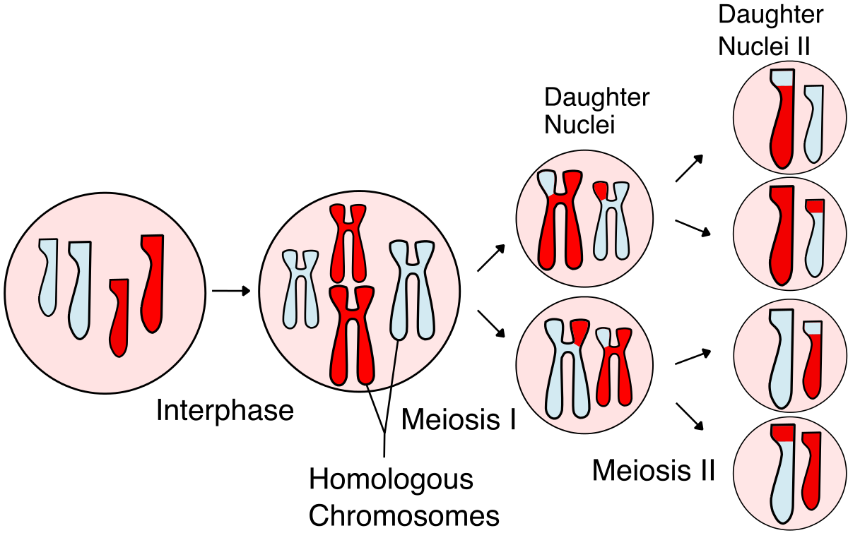
One kind of reassortment is a consequence of the random distribution of the maternal and paternal homologs between the daughter cells at meiotic division I, as a result of which each acquires a different mixture of maternal and paternal chromosomes. From this process alone, one individual could, in principle, produce 2n genetically different gametes, where n is the number of chromosomes . In humans, for example, each individual can produce at least 223 = 8.4 × 10⁶ genetically different gametes. But the actual number of variants is very much greater than this because a second type of reassortment, called , occurs during . It takes place during the long of meiotic division I , in which parts of chromosomes are exchanged. On average, between two and three crossover events occur on each pair of human chromosomes during meiotic division I. This process scrambles the genetic constitution of each of the chromosomes in gametes, as illustrated in .
Two major contributions to the reassortment of genetic material that occurs in the production of gametes during meiosis. The independent assortment of the maternal and paternal homologs during the first meiotic division produces 2n different haploid
Changing Partners At The Dance

The structure and function of the SC is conserved in sexually reproducing organisms , and influences the most critical outcome of meiotic prophase Ithe exchange of DNA between maternal and paternal chromosomes or CO. Because COs occur only in the context of assembled CEs, the SC is thought to be a major factor in CO regulation, although how this regulation is exerted is only partially understood. Crossovers are tightly controlled: most organisms have at least one obligatory CO per homolog pair COs are nonrandomly positioned, so that adjacent COs are further apart than expected if they were randomly distributed and total CO numbers are maintained at a relatively constant level . Interestingly, class II COs are not constrained by interference and it remains unclear whether they are regulated by homeostatic mechanisms .
Mechanisms underlying CO assurance have been difficult to study, and involve processes of both SC assembly and recombination. Interestingly, in oocytes lacking AE proteins SYCP2/3, COs are formed and interference is maintained in spite of severely compromised SC structure , supporting the idea that the mechanisms regulating CO numbers and distribution are established before SC is formed . In mice, any perturbation of CE assembly impairs DSB repair and leads to meiotic arrest before CO can be observed .
Also Check: Child Of Rage Brother Jonathan Now
Comparison Ofmitosis And Meiosis
Mitosis maintains ploidy level, while meiosisreduces it. Meiosis may be considered a reduction phase followed by aslightly altered mitosis. Meiosis occurs in a relative few cells of amulticellular organism, while mitosis is more common.
Comparison of the events in Mitosis andMeiosis. Images from Purves et al., Life: The Science ofBiology, 4th Edition, by Sinauer Associates and WH Freeman ,used with permission.
Chiasmata Have An Important Role In Chromosome Segregation In Meiosis
In addition to reassorting genes, is crucial in most organisms for the correct segregation of the two duplicated homologs to separate daughter nuclei. This is because the chiasmata created by crossover events have a crucial role in holding the maternal and paternal homologs together until the spindle separates them at I . Before anaphase I, the two poles of the spindle pull on the duplicated homologs in opposite directions, and the chiasmata resist this pulling. In organisms that have a reduced frequency of meiotic crossing-over, some of the chromosome pairs lack chiasmata. These pairs fail to segregate normally, and many of the resulting gametes contain too many or too few chromosomes.
The duplicated homologs are held together at chiasmata only because the arms of sister chromatids are glued together along their length by proteins called cohesins . In Drosophila, for example, if a -specific cohesin is defective, sister chromatids separate prior to I and, as a consequence, the homologs segregate abnormally.
As illustrated in , the arms of sister chromatids suddenly become unglued at the start of I, when the cohesins holding the arms together are degraded, allowing the duplicated homologs to separate and be pulled to opposite poles of the spindle. The sister chromatids of each duplicated remain attached at the by -specific cohesins, which are degraded at anaphase of meiotic division II only then can the sister chromatids separate.
Don’t Miss: How To Find Delta Math Answers With Inspect Element
Age Effects In Meiosis
Both sexual dimorphism in meiosis and the diversification of gametogenesis programs that arose during evolution pose problems for human reproduction. Females appear to have chosen to guard genome quality by limiting the number of cell divisions, thus decreasing the risk of potential de novo germline mutations. The downside is that they produce fewer eggs, and these have to be stored for a long time before they can be fertilized. This strategy may not be a problem when females produce offspring at a young age, but this is not always the case in the modern human population. The strategy in males appears to be to continuously produce large quantities of gametes from continually rejuvenating precursors however, this potentially sacrifices gametic genomic quality due to mitotic mutation rate and consequent age-related accumulation of de novo germline mutations . Likewise, this can be an issue among human males extending fatherhood to an advanced age.
Meiosis In Humans And Other Animals
How does meiosis work in humans? Meiosis produces haploid gametes in humans and other animals. It is a crucial part of gametogenesis. As the name implies, gametogenesis is the biological process of creating gametes. In humans and other animals, there are two forms of gametogenesis: spermatogenesis and oogenesis .
In oogenesis, four haploid gamete cells are produced from a diploid oocyte. However, only one cell survives and functions as an egg the other three become polar bodies. This effect results from the unequal division of the oocyte by meiosis where one of the formed cells receives most of the cytoplasm of the parent cell while the other formed cells degenerate which contributes to increasing the concentration of the nutrients in the formed egg. The egg cell acquires most of its specialized functions during phases of meiosis especially prophase I.
In spermatogenesis, the sperm acquires its specialized features in order to develop into a functional gamete after meiosis and post-meiotic events, e.g. spermiogenesis where the sperm cell matures by acquiring a functional flagellum and discarding most of their cytoplasm to form a compacted head.
Read Also: Beth Thomas Father Jailed
Other Types Of Trisomy
Most cases of trisomy result in miscarriage during the first trimester of pregnancy because the fetus cannot survive the chromosomal abnormality. Trisomy 16 occurs in over 1% of pregnancies and is the most common trisomy, but most individuals with this trisomy do not survive unless some of their cells are normal.
The three most common types of trisomy that are survivable are Trisomy 21 , Trisomy 18 , and Trisomy 13 .
The reason these chromosomal abnormalities are more common is due to the specific chromosomes they affect. Chromosome 21, 18, and 13 are all relatively small and gene-poor they dont have as many genes on them as other chromosomes. This means they have less of a drastic effect on the cell. As a result, more individuals survive with these trisomies. Others are non-viable.
In Plants And Animals
Meiosis occurs in all animals and plants. The end result, the production of gametes with half the number of chromosomes as the parent cell, is the same, but the detailed process is different. In animals, meiosis produces gametes directly. In land plants and some algae, there is an alternation of generations such that meiosis in the diploid sporophyte generation produces haploid spores. These spores multiply by mitosis, developing into the haploid gametophyte generation, which then gives rise to gametes directly . In both animals and plants, the final stage is for the gametes to fuse, restoring the original number of chromosomes.
Don’t Miss: Oxygen Difluoride Polar Or Nonpolar
B Phases Of Meiosis Ii
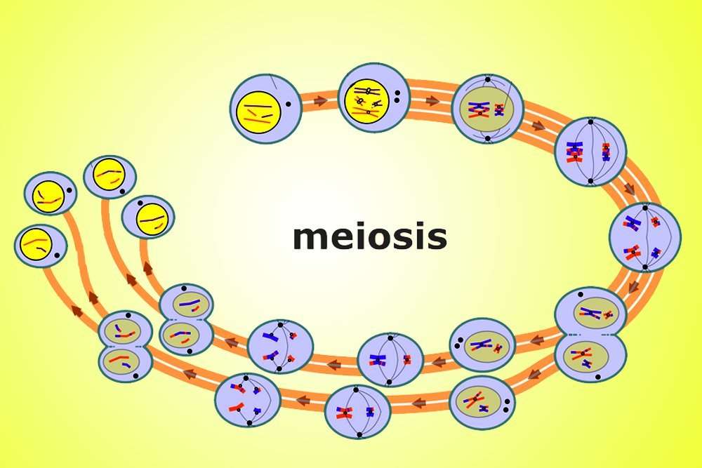
Interphase meiosis begins after the end of meiosis I and before the beginning of meiosis II, this stage is not associated with the replication of DNA since each chromosome already consists of two chromatids that were replicated already before the initiation of meiosis I by the DNA synthesis process. In brief, DNA is replicated before meiosis I start at one time only. The stage of meiosis II or second mitotic division has a purpose similar to that of mitosis where the two new chromatids are oriented in two new daughter cells. Therefore, the second meiotic division is sometimes referred to as .
Step 1: Prophase II
Prophase 2 is the stage that follows meiosis I or interkinesis, it is characterized by the nuclear envelope and nucleolus disintegration as well as the chromatids thickening and shortening in prophase II, and centrosomes replicate and migrate to the polar side. Prophase II is simpler and shorter than prophase I it somehow resembles the mitotic prophase. On the other hand, prophase II is different from prophase I since crossing over of chromosomes occurs during prophase I only and not prophase II. Metaphase II starts at the end of prophase II.
Step 2: Metaphase II
Metaphase 2 of meiotic division is also similar to metaphase of mitotic division, however, only half the number of chromosomes are present in metaphase II, metaphase II is characterized by the chromosomal alignment in the center of the cell.
Step 3: Anaphase II
Step 4: Telophase II
Results of meiosis II
Meiotic Chromosome Pairing Culminates In The Formation Of The Synaptonemal Complex

A series of events occurs during the long of meiotic division I: duplicated chromosomes pair, is initiated between nonsister chromatids, and each pair of duplicated homologs assembles into an elaborate structure called the . In some organisms, genetic recombination begins before the assembles and is required for the complex to form in others, the complex can form in the absence of recombination. In all organisms, however, the recombination process is completed while the is held in the synaptonemal complex, which serves to space out the crossover events along each .
The of meiotic division I is traditionally divided into five sequential stages, , , , and diakinesisdefined by the morphological changes associated with the assembly and disassembly of the . Prophase begins with , when the duplicated paired homologs condense. At , the synaptonemal complex begins to develop between the two sets of sister chromatids in each . begins when synapsis is complete, and it generally persists for days, until desynapsis begins the stage, in which the chiasmata are first seen .
Chromosome synapsis and desynapsis during the different stages of meiotic prophase I. A single bivalent is shown. The pachytene stage is defined as the period during which a fully formed synaptonemal complex exists. At leptotene, the two sister chromatids
Recommended Reading: Paris Jackson Real Father
How Do Cells Divide
There are two types of cell division: mitosis and meiosis. Most of the time when people refer to cell division, they mean mitosis, the process of making new body cells. Meiosis is the type of cell division that creates egg and sperm cells.
Mitosis is a fundamental process for life. During mitosis, a cell duplicates all of its contents, including its chromosomes, and splits to form two identical daughter cells. Because this process is so critical, the steps of mitosis are carefully controlled by certain genes. When mitosis is not regulated correctly, health problems such as cancer can result.
The other type of cell division, meiosis, ensures that humans have the same number of chromosomes in each generation. It is a two-step process that reduces the chromosome number by halffrom 46 to 23to form sperm and egg cells. When the sperm and egg cells unite at conception, each contributes 23 chromosomes so the resulting embryo will have the usual 46. Meiosis also allows genetic variation through a process of gene shuffling while the cells are dividing.
Mitosis and meiosis, the two types of cell division.
Genetic Maps Reveal Favored Sites For Crossovers
Using such measurements, geneticists have constructed detailed of the entire human , in which the distance between each pair of neighboring genes is displayed as the percentage between them. The standard unit of genetic distance is the , which corresponds to a 1% probability that two genes will be separated by a crossover event during . A typical human is more than 100 centimorgans long, indicating that more than one crossover is likely to occur on a typical human chromosome.
Another way to construct a is to measure the coinheritance of short sequences that differ between individuals in the populationthat is, that are . Genetic maps constructed in this way have two advantages over genetic maps constructed by tracing the phenotypes of individuals that inherit genes. First, they can be more detailed, as there are large numbers of DNA markers that can be measured. Second, they can reveal the real distance in pairs between the markers, so that genetic distances in centimorgans can be compared directly with true physical distances along a .
Comparison of the physical and genetic maps of part of chromosome I in budding yeast. The DNA markers shown are various genes. A indicates a region where the genetic map is contracted owing to decreased frequency of crossing-over. B indicates a region
Don’t Miss: Span Definition Linear Algebra
The Phases Of Mitosis
Prophase
The first phase of mitosis is prophase. During prophase, the cells chromosomes condense and become visible under a light microscope. The nucleolus disappears, and the mitotic spindle begins to form.
Prometaphase
The nuclear membranebreaks down. The microtubules attach themselves to the chromosomes and begin to move them around.
Metaphase
The microtubules move the chromosomes until they are lined up along the middle of the cell. This line of chromosomes is called the metaphase plate.
Anaphase
The chromosomes are pulled apart by the microtubules. Each chromosome is separated into two, genetically identical sister chromatids, which are pulled to opposite ends of the cell.
Telophase
The sister chromatids arrive at opposite ends of the cell. A new nuclear membrane begins to form around each set of chromosomes. The chromosomes so they are no longer visible under a light microscope. The nucleolus reappears, and the mitotic spindle disappears.
Finally, the cytoplasm of the cell splits, and two new, genetically identical daughter cells are formed. This process is called cytokinesisand usually takes place during telophase.
Early Prophase I Of Meiosis
Much like mitosis, Early Prophase I of meiosis begins with chromosomes condensing and the nuclear envelope beginning to disintegrate. Unlike prophase of mitosis, homologous chromosome pairs come together forming a tetrad, in which the four chromatids are connected along the length of each chromatid. Chromatids from different homologous pairs are referred to as non-sister chromatids.
Don’t Miss: Define Movement In Geography
Nondisjunction During Meiosis Ii
Even if a cell divides normally in meiosis I, nondisjunction can still occur in the second round of meiosis, meiosis II. During meiosis II, each of the two daughter cells produced during meiosis I undergo a second round of meiosis, going from diploid to haploid . This allows them to participate in sexual reproduction once the egg and sperm combine during fertilization, the diploid state is restored.
If a pair of sister chromatids fail to separate properly during anaphase of meiosis II, one daughter cell will have an extra chromosome and one daughter cell will be missing a chromosome. If the other daughter cell created in meiosis I splits properly, the other two of the four total daughter cells created during meiosis II will have the normal number of chromosomes.
Thus, the effects of nondisjunction during meiosis are observed only in the offspring of the individual. The effects of non-disjunction during mitosis are only observed in that individual and are not passed on to the next generation.